Best known for his world-renowned neuro-ophthalmology unit based at the University of California, San Francisco, William Hoyt, MD collected here more than 850 of his best images covering a wide range of disorders.
William F. Hoyt, MD, Professor Emeritus of Ophthalmology, Neurology and Neurosurgery, Department of Ophthalmology, University of California, San Francisco.
NOVEL: https://novel.utah.edu/
TO
| Title | Description | Type | ||
|---|---|---|---|---|
| 1 |
 |
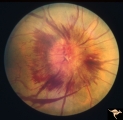
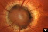
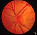
B301 Disc Swelling, Giant Cell Arteritis | Disc swelling. Giant Cell Arteritis. Temporal. Ischemic swelling. Blind eye with pallid swelling and marked dilation of central retinal vein. | Image |
| 2 |
 |
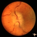
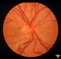
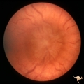
Bilateral Crowded Discs | Left eye. Bilateral crowded discs with congenital blurring. Blurred disc margins are not from edema. Note optic cup is absent. Pair with right eye in PP_1a, and brother in PP_2. Mother has drusen of the optic disc in PP_11aa & b. Sister has drusen in PP_11c. Anatomy: Optic disc. Pathology: Normal va... | Image |
| 3 |
 |
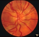
Bilateral Crowded Discs (Family) | Right eye. Bilateral crowded discs with congenital blurring. Blurred disc margins are not from edema. Note optic cup is absent. Pair with left eye in PP_1b, and brother in PP_2. Mother has drusen of the optic disc in PP_11a & b. Sister has drusen in PP_11c. Anatomy: Optic disc. Pathology: Normal var... | Image |
| 4 |
 |
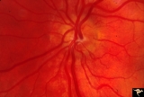
Bilateral Hemorrhagic Papilledema | Left eye. Bilateral Hemorrhagic Papilledema from cardio-respiratory disease. Woman. Anatomy: Optic disc. Pathology: Bilateral papilledema, hemorrhagic. Disease/Diagnosis: Pseudotumor due to cardio-respiratory disease. Clinical notes: Woman with headache, shortness of breath. | Image |
| 5 |
 |
Bilateral Hemorrhagic Papilledema | Bilateral Hemorrhagic Papilledema from cardio-respiratory disease. Woman. Anatomy: Optic disc. Pathology: Bilateral papilledema, hemorrhagic. Disease/Diagnosis: Pseudotumor due to cardio-respiratory disease. Clinical notes: Woman with headache, shortness of breath. | Image |
| 6 |
 |
Bilateral Severe Hemorrhagic Papilledema | Left eye. Two months later, resolving Bilateral Severe Hemorrhagic Papilledema. Same eye as P_32b | Image |
| 7 |
 |
Bilateral Severe Hemorrhagic Papilledema | Right eye. 2 months later, resolving Bilateral Severe Hemorrhagic Papilledema. Same eye as P_32a | Image |
| 8 |
 |
Bilateral Severe Hemorrhagic Papilledema | Right eye. Bilateral Severe Hemorrhagic Papilledema in a woman with hyperthyroidism and dural sinus occlusion. | Image |
| 9 |
 |
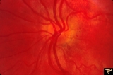
Bilateral Severe Hemorrhagic Papilledema | Left eye. Bilateral Severe Hemorrhagic Papilledema in a woman with hyperthyroidism and dural sinus occlusion. | Image |
| 10 |
 |
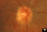
Buried Drusen | Buried drusen with peculiar white dot, which appears to be choroidal in location. Note lumpy disc margin on right disc PP_15a is right eye. PP_15b is left eye. Beautiful example of pseudo papilledema in which drusen can not be seen. 8 year old girl. Anatomy: Optic disc. Pathology: Drusen of the op... | Image |
| 11 |
 |
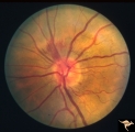
Buried Drusen | Left disc has a blurred lumpy margin. Retinal vessels are not obscured in the disc margin blur, therefore no edema is present. This is an example of a difficult blurred disc, the nature of which is clarified by the presence of a clear cut disk anomoly in the fellow eye. 8 year old girl. PP_15a has b... | Image |
| 12 |
 |
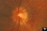
C14 Morning Glory Disc | Isolated "Morning Glory". Left eye. Girl. Anatomy: Optic disc. | Image |
| 13 |
 |
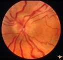
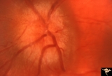
Crowded Disc (Family) | Right eye. PP3 a & b: sister; PP4 a & b brother; Congenital disc margin blurring with crowded discs. Excellent example of pseudo papilledema. Anatomy: Optic disc. Pathology: Normal variant of the optic disc. Disease/Diagnosis: Normal variant of the optic disc. Crowded disc. Clinical: Appearance is ... | Image |
| 14 |
 |
Crowded Disc (Family) | Right eye. PP3 a & b: sister; PP4 a & b brother; Congenital disc margin blurring with crowded discs. Excellent example of pseudo papilledema that caused serious diagnostic confusion which led to a pneumoencephalogram (PEG) and arteriogram. Anatomy: Optic disc. Pathology: Normal variation of the opt... | Image |
| 15 |
 |
Crowded Disc (Family) | Left eye. PP3 a & a: sister; PP4 a & b brother; Congenital disc margin blurring with crowded discs. Excellent example of pseudo papilledema that caused serious diagnostic confusion which led to a pneumoencephalogram (PEG) and arteriogram. Anatomy: Optic disc. Pathology: Normal variation of the opti... | Image |
| 16 |
 |
Crowded Disc (Family) | Left eye. PP3 a & b: sister; PP4 a & b brother; Congenital disc margin blurring with crowded discs. Excellent example of pseudo papilledema. Anatomy: Optic disc. Pathology: Normal variation of the optic disc. Disease/Diagnosis: Normal variation of the optic disc. Crowded disc. Clinical: Appearance ... | Image |
| 17 |
 |
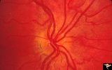
Crowded Disc (Family) | Anomalous vasculature with congenital disc margin blurring. Note optic cup is absent. Pair with brother in PP1a & b. Mother has drusen of the optic disc in PP11aa & b. Sister has drusen in PP11c. Anatomy: Optic disc. Pathology: Normal variant. Cause of appearance is too many fibers entering into a s... | Image |
| 18 |
 |
F105 Histiocytosis Infiltrate of Disc | Histiocytosis infiltrate of right disc with simultaneous infiltration of the hypothalamus with skin lesions on eye lids and chest. Same patient as F1_06. Anatomy: Optic disc. Pathology: Histiocytosis infiltrate. Disease/ Diagnosis: Histiocytosis infiltrate. Clinical: Patient presented with skin lesi... | Image |
| 19 |
 |
F2b01 Optic Nerve Glioma | Left eye. Woman with optic nerve glioma. Anatomy: Optic disc. Pathology: Optic nerve swelling secondary to retrobulbar optic glioma. Disease/ Diagnosis: Optic nerve glioma. | Image |
| 20 |
 |
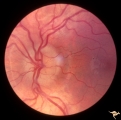
Familial Drusen | Right eye: Mother with obvious optic nerve drusen. Note the blurred temporal margin where buried drusen can not be seen.; PP_11b: mother visible drusen; Buried drusen; lumpy disc. Combine with PP_1a & b and PP_2 (sons) and PP_11c (daughter). Anatomy: Optic disc. Pathology: Drusen of the optic dis... | Image |
| 21 |
 |
Familial Drusen | Left eye. PP_11b: Mother visible drusen; buried drusen; lumpy disc. PP_11a: Mother with obvious optic nerve drusen; Combine with PP_1a & b and PP_2 (sons) and PP_11c (daughter). Anatomy: Optic disc. Pathology: Drusen of the optic disc. Disease/Diagnosis: Drusen of the optic disc. Clinical: Congen... | Image |
| 22 |
 |
Familial Drusen | PP11c: daughter: buried drusen; lumpy disc. Combine with PP1a & b and PP2 (brothers) and PP11a & b (mother). Anatomy: Optic disc. Pathology: Drusen of the optic disc. Disease/Diagnosis: Drusen of the optic disc. Clinical: Congenital dominant hereditary drusen. | Image |
| 23 |
 |
H09 Panhypoplasia | Moderate hypoplasia. Man. Anatomy: Optic disc. Pathology: Hypoplasia of the optic nerve. Disease/ Diagnosis: Hypoplasia. | Image |
| 24 |
 |
H23 Dysplasia with Hypoplasia (Elevated Hysplasia with Anomalous Vessels) | Elevated dysplasia with anomalous vessels. Left eye. Hypoplasia with central glial tissue remnant. Japanese girl. Same patient as H_24. Anatomy: Optic disc. Pathology: Dysplasia of the optic disc. Disease/ Diagnosis: Elevated dysplasia with hypoplasia. | Image |
| 25 |
 |
H24 Dysplasia with Hypoplasia (Elevated Dysplasia with Anomalous Hessels) | Elevated dysplasia with anomalous vessels. Right eye. Hypoplastic with dysplasia. Japanese girl. Same patient as H_23. Anatomy: Optic disc. Pathology: Dysplasia of the optic disc. Disease/ Diagnosis: Elevated dysplasia with hypoplasia. | Image |