Collection of materials relating to neuro-ophthalmology as part of the Neuro-Ophthalmology Virtual Education Library.
NOVEL: https://novel.utah.edu/
TO
- NOVEL978
Filters: Collection: "ehsl_novel_novel"
| Title | Creator | Description | Subject | ||
|---|---|---|---|---|---|
| 226 |
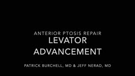 |
Anterior Ptosis | Patrick Burchell, MD; Jeffrey Nerad, MD | Demonstration of anterior ptosis repair, levator advancement. | Anterior Ptosis |
| 227 |
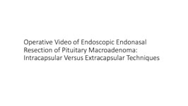 |
Pituitary Surgery | Jonathan Forbes | Operative video of endoscopic endonasal resection of pituitary macroadenoma. Describes intracapsular versus extracapsular techniques. | Pituitary Surgery |
| 228 |
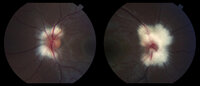 |
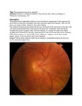
Peripapillary Myelinated Nerve Fibers | John J. Chen, MD, PhD | Fundus photographs of a 19-year old female with prominent peripapillary myelinated nerve fibers in both eyes that was incidentally found on routine eye examination. | Myelinated Nerve Fibers |
| 229 |
|
Myelinated Nerve Fibers | John J. Chen, MD, PhD | Fundus photographs of a 19-year old female with prominent peripapillary myelinated nerve fibers in both eyes that was incidentally found on routine eye examination. | Myelinated Nerve Fibers |
| 230 |
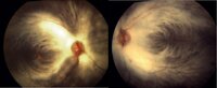 |
Myelinated Nerve Fibers | Carmen Chan,RN, PhD, FAAN | Fundus photos from a patient with extensive myelinated nerve fibers. The patient had normal visual functions. | Myelinated Nerve Fibers |
| 231 |
 |
Common Vitreo-Retinal Procedures and Surgeries | Luke B. Lindsell, OD, MD | Brief presentation on Common vitreo-retinal procedures and surgeries. | Vitreo-Retinal Procedures; Surgeries |
| 232 |
 |
Myotonic Dystrophy | Brian Villafuerte, MD, Ezequiel Piccione, MD | Presentation covering an overview of myotonic dystrophy. | Myotonic Dystrophy |
| 233 |
 |
Congenital Hydrocephalus | Mays El-Dairi, MD | Presentation covering an overview of congenital hydrocephalus. | Congenital Hydrocephalus |
| 234 |
 |
Fibrous Dysplasia | Mays El-Dairi, MD | Presentation covering an overview of fibrous dysplasia. | Fibrous Dysplasia |
| 235 |
 |
Optic Neuropathy: A Recipe for Blindness | Karim Kozhaya, MD; Alaa Bou Ghannam, MD; Alfredo Sadun, MD, PhD | An epidemic of blindness and peripheral neuropathy struck Cuba in the early 90s. By the end of 1993, 7% of the population was affected. Most patients were men and presented with sub-acute, painless, bilateral loss of vision. The etiology of the disease pondered local and international scientists, es... | Cuban Epidemic Optic Neuropathy; Leber's Hereditary Optic Neuropathy; Mitochondrial Insufficiency; Nutritional Optic Neuropathy; Pale Optic Nerve |
| 236 |
 |
Heavy Eye Syndrome | Meagan D. Seay, DO; Bradley J. Katz, MD | A brief overview of heavy eye syndrome. | Heavy Eye Syndrome |
| 237 |
 |
Brown Syndrome | Meagan Seay, DO | A brief overview of Brown Syndrome. | Brown Syndrome |
| 238 |
 |
Intraocular Pressure (IOP) Measurement: A Simple Guide | Nandini Singh; Kirsty Sumerville Mcalester; Alicia Yap; Anne Lee | This video demonstrates the technique for measuring intraocular pressure (IOP) and the use of the tonopen. | Intraocular Pressure (IOP); Tonometry |
| 239 |
 |
Retinal Detachment: The Basics | Arnav Gupta, BHSc; Rahul Sharma, MD, MPH | A presentation describing retinal detachment. | Retinal Detachment |
| 240 |
 |
Retinal Exudate: The Basics | Arnav Gupta, BHSc; Rahul Sharma, MD, MPH | A presentation describing retinal exudate. | Retinal Exudate |
| 241 |
 |
Retinal Hemorrhage: The Basics | Arnav Gupta, BHSc; Rahul Sharma, MD, MPH | A presentation describing retinal hemorrhage. | Retinal Hemorrhage |
| 242 |
 |
Introduction to the Evaluation of Visual Function in NANOS NOTE | Sean Gratton, MD | Introduction to the Evaluation of Visual Function in NANOS NOTE | Visual Function; Examination |
| 243 |
 |
Situs Inversus Optic Disc Anomaly | Michael Hii, Medical Student; Ryan Walsh, MD | This patient was incidentally-noted to have anomalous appearance of the optic discs, right more so than left, consistent with situs inversus optic disc anomaly. She did not have any visual deficits related to this exam finding. ; The patient's fundus photos demonstrate situs inversus of the optic ... | Situs Inversus Optic Disc Anomaly |
| 244 |
 |
Temporal Artery Biopsy Procedure | Nooran Badeeb; Danah Albreiki | Temporal artery biopsy is a procedure that is done in a patient with suspicion of GCA (Giant cell Arteritis), and some of the clinical manifestations that prompts us to suspect the diagnosis in patients older than 50 years old are: 1. GCA symptoms e.g. new onset headache. 2 . Visual symptoms: - Visi... | Temporal Artery Biopsy; GCA; Temporal Arteritis |
| 245 |
 |
An Introduction to CT and MRI in Neuro-Imaging | Michael Carper, MD | A brief lecture covering basic neuro-imaging, including computed tomography (CT) and magnetic resonance imaging (MRI). | Computed Tomography (CT); Magnetic Resonance Imaging (MRI); Neuro-imaging |
| 246 |
 |
Age-related Macular Degeneration: The Basics | Arnav Gupta, BHSc; Rahul Sharma, MD, MPH | A presentation covering age-related macular degeneration ("ARMD" or "AMD"), an acquired, progressive, chronic, degenerative disease of the retina. | Macular Degeneration |
| 247 |
 |
Night Wolf | Mehdi Tavakoli, MD; Byron Lam, MD | A case presentation on radiation optic neurology. | Radiation Optic Neuropathy |
| 248 |
 |
Muscular Dystrophy Classification | Brian Villafuerte, MD, Ezequiel Piccione, MD | Presentation covering an overview of muscular dystrophy classification. | Muscular Dystrophy Classification |
| 249 |
 |
Radiation Optic Neuropathy | Neil R. Miller, MD, FACS | Overview of Radiation Optic Neuropathy (RON). | Radiation Optic Neuropathy; RON |
| 250 |
 |
Tolosa Hunt Syndrome | Sahil Aggarwal, MD; Jason Liss, MD | Presentation covering an overview of Tolosa Hunt Syndrome. | Tolosa Hunt Syndrome |