Collection of materials relating to neuro-ophthalmology as part of the Neuro-Ophthalmology Virtual Education Library.
NOVEL: https://novel.utah.edu/
TO
- NOVEL970
Filters: Collection: "ehsl_novel_novel"
| Title | Creator | Description | Subject | ||
|---|---|---|---|---|---|
| 26 |
 |
Trigeminal Autonomic Cephalgias | Kaitlin Keenan; Aimee J. Szewka | These slides are a comprehensive discussion addressing Trigeminal Augonomic Cephalgias - in particular, Cluster Headaches, Paroxysmal Hemicrania, Hemicrania Continua and Short-Lasting Unilateral Neuralgiform Headache Attack (SUNHA). Our slides discuss diagnosis, prognosis and treatment of these rare... | Cluster Headache; Headache; Hemicrania Continua; Paroxysmal Hemicrania; Short-Lasting Unilateral Neuralgiform Headache Attack; Trigeminal Autonomic Cephalgia |
| 27 |
 |
Neuro-ophthalmologic Findings in Cerebellar Stroke | Zoe Rebecca Williams; Aishwarya Ganesh | This PowerPoint reviews the anticipated neuro-ophthalmologic findings in cerebellar strokes including posterior inferior cerebellar artery (PICA), anterior inferior cerebellar artery (AICA) and superior cerebellar artery (SCA) territory infarcts. The review includes discussion of potential ocular mi... | AICA; Cerebellar Esotropia; Cerebellar Infarct; Lateral Medullary Syndrome; Nystagmus; Ocular Lateropulsion; PICA; SCA; Skew Deviation |
| 28 |
 |
Gustatory Lacrimation: Crocodile Tear Syndrome | Fernando Pellerano; Kevin Lai | We describe a case of gustatory lacrimation on a 61-year-old male with a history of Bell's Palsy that presented to the Ophthalmology clinic with painless epiphora in the right eye while eating. Examination was notable for oral-ocular synkinesis and hyperlacrimation while eating on the right eye only... | Crocodile Tear Syndrome; Gustatory Lacrimation; Oral-ocular Synkinesis |
| 29 |
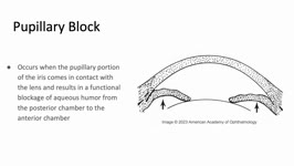 |
Pupillary Block | Sujata Dalal; James Brian Davis; Amanda Dean Henderson | Pupillary block occurs when the pupillary margin of the iris contacts the anterior surface of the lens. This creates a barrier for the outflow of aqueous humor from the posterior chamber to the anterior chamber and resultant increased pressure in the posterior chamber. This pressure can cause the ir... | Angle-Closure Glaucoma; Iris Bombe; Narrow Angle; Pupillary Block |
| 30 |
 |
Behind the Mask | Pame Dávila; Nagham Al-Zubidi | Case report describing temporal artery amyloidosis masquerading as giant cell arteritis. | Giant cell arteritis; Temporal arteritis; Temporal artery amyloidosis |
| 31 |
 |
Reversible Cerebral Vasoconstriction Syndrome (RCVS) | Felix Yang; Sean Gratton | This is a narrated powerpoint that reviews Reversible Cerebral Vasoconstriction syndrome. It discusses the diagnostic basics and evaluation and management. It compares RCVS to Posterior Reversible Encephalopathy Syndrome (PRES). | Primary Angiitis of the Central Nervous System; Reversible Cerebral Vasoconstriction Syndrome; Subarachnoid Hemorrhage; Thunderclap Headache |
| 32 |
 |
Glaucoma: Evaluation and Principles of Management | Anand Haran; Sean Gratton | This is a narrated powerpoint that reviews the basics of glaucoma. It covers Primary Open Angle Glaucoma and Angle Closure Glaucoma, and includes reviews of the pathophysiology, evaluation, and management | Angle Closure Glaucoma; Glaucoma; Primary Open Angle Glaucoma |
| 33 |
 |
Idocyanine Green Angiography | Tanner Nordseth; Evan Wotipka; Talhah Zubair; Anne Abel; Hossein Nazari | This narrated PowerPoint presentation gives an overview of idocyanine green angiography. Historical significance, diagnostic utility, and comparison to fluorescein angiography are discussed. | Angiography; Choroidal imaging; Idocyanine green |
| 34 |
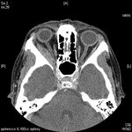 |
Sixth nerve palsy or not? (Image) | Vivian Paraskevi Douglas; Konstantinos Douglas; Nurhan Torun | Here in we present a case of a 72-yearold Caucasian female with remote history of breast cancer who presented with diplopia in the right gaze over the past several months. There had been no improvement after a 5-day course of steroids by an outside ophthalmologist. On neuro-ophthalmic examination, t... | Abduction deficit; Diplopia; Orbital imaging |
| 35 |
 |
Sixth nerve palsy or not? (Video) | Vivian Paraskevi Douglas; Konstantinos Douglas; Nurhan Torun | Here in we present a case of a 72-yearold Caucasian female with remote history of breast cancer who presented with diplopia in the right gaze over the past several months. There had been no improvement after a 5-day course of steroids by an outside ophthalmologist. On neuro-ophthalmic examination, t... | Abduction deficit; Diplopia; Orbital imaging |
| 36 |
 |
Diagnosis and Evaluation of Stroke: Cerebral Hypoxia | Danny Alevy; James Brian Davis; Amanda Dean Henderson | Neurons are particularly vulnerable to oxygen deprivation. Cerebral hypoxia can be caused by arterial thrombosis, embolism, hypoperfusion, cervical artery dissection, or cryptogenic causes. About 1/3 of ischemic strokes are from cryptogenic causes. Embolic strokes are the next most common (20-30%), ... | Atherosclerosis; Cerebral Hypoxia; Cervical Artery Dissection; Cryptogenic Ischemia; Embolism; Hypoperfusion; Ischemia; Stroke; Thrombosis |
| 37 |
 |
One and a Half Syndrome Following Resection of a Posterior Fossa Epidermoid Cyst | Christine Xu; Claire Basco, MS, NP-C; Kiarash Shahlaie; Yin Allison Liu | This is a case of One and a Half Syndrome following resection of a posterior fossa epidermoid cyst. A 31-year-old male initially presented with left facial droop and bilateral ptosis, and a "down and out" gaze of the left eye. He underwent imaging and was diagnosed with an epidermoid cyst located in... | Abducens Nucleus; Epidermoid Cyst; Extraocular Movements; Gaze Palsy; Internuclear Ophthalmoplegia; Medial Longitudinal Fasciculus; Neurosurgery; One and a Half Syndrome |
| 38 |
 |
Neuro-Ophthalmic Complications of Chemotherapy | Nagham Al-Zubidi; May Ameri | This lecture covers the effect of antineoplastic chemotherapy, which can cause damage to the optic nerve and the ocular motor nerves. | Antineoplastic Chemotherapy; Ocular Motor Nerve Damage; Optic Atrophy; Optic Nerve Damage; Optic Nerve Edema; Optic Neuritis |
| 39 |
 |
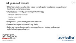
Giant Cell Arteritis with Choroidal Infarction | Aretha Zhu; Kareem Moussa; Tina Zeng; Yin Allison Liu | The presentation describing a case of giant cell arteritis (GCA) with choroidal infarction documents an 83-year-old male experiencing recurring episodes of vision loss, predominantly manifesting as a "graying" of vision in both eyes, associated with new-onset headaches. The individual presented to t... | Choroidal Infarction; Erythrocyte Sedimentation Rate, Giant Cell Arteritis; Headache; Jaw Claudication; Painless Vision Loss; Rheumatology; Temporal Artery Biopsy; Vasculitis |
| 40 |
 |
Patient Portal: Optic Disc Drusen | Cristiano Oliveira, MD | Optic disc drusen (ODD) are abnormal deposits of benign, usually calcified material within the optic disc, which is the front part of the optic nerve that connects each eye to the brain. We do not know the exact cause of optic disc drusen. They are present in 0.3-2% of people as an isolated case or ... | Optic disc drusen; Papilledema; Pseudopapilledema |
| 41 |
 |
Patient Portal: Microvascular Cranial Nerve Palsy | Meagan Seay, DO | A nerve palsy is an impairment in the function of a nerve, which results in a decrease in function of the corresponding muscles controlled by that nerve. In microvascular cranial nerve palsy, something affects the blood supply to one of the cranial nerves, causing it not to work. This is usually the... | Nerve palsy; Microvascular cranial nerve palsy; Cranial nerve 3; CN3; Oculomotor nerve; Cranial nerve 4; CN4; Trochlear nerve; Cranial nerve 6; CN6; Abducens nerve |
| 42 |
 |
Patient Portal: Non-Arteritic-Anterior Ischemic Optic Neuropathy (NAION) | Arun Sundaram, MD | Non-arteritic anterior ischemic optic neuropathy (NAION or NA-AION) is caused by decreased blood flow to the front part of the optic nerve (optic disc). It causes optic nerve swelling and sudden vision loss. NAION typically affects one eye, although the other eye sometimes suffers similar loss month... | Non-arteritic anterior ischemic optic neuropathy; NAION; NA-AION; Optic nerve; Optic disc; Ophthalmic artery |
| 43 |
 |
Patient Portal: Myasthenia Gravis | Aroucha Vickers, DO | Myasthenia gravis (MG) is an autoimmune disease in which the body's immune system creates antibodies (proteins that normally protect us) that may attack receptors on your muscles. This results in muscle weakness because the muscles do not receive the signals to contract (tighten). Muscles anywhere w... | Myasthenia gravis; Ptosis; Double vision |
| 44 |
 |
Patient Portal: Giant Cell Arteritis | Anne S. Abel, MD | Giant cell arteritis is an inflammatory condition that can cause vision loss, double vision, fever, new persistent headaches, scalp tenderness, and jaw pain with chewing. GCA is caused by inflammation of blood vessels, primarily in the head and neck. Sometimes called "temporal arteritis," GCA frequ... | Giant cell arteritis |
| 45 |
 |
Patient Portal: Idiopathic Intracranial Hypertension (IIH) | Devin D. Mackay, MD | Idiopathic intracranial hypertension (IIH), also called pseudotumor cerebri, is a condition in which there is high pressure in the fluid surrounding your brain, spinal cord and optic nerves. This can cause headaches and problems with vision. Although the cause(s) of the condition is not fully unders... | Idiopathic intracranial hypertension; Pseudotumor cerebri |
| 46 |
 |
Patient Portal: Homonymous Hemianopsia | James C. O'Brien, MD | Homonymous hemianopia refers to an absence of vision towards one side of the visual world in each eye. The damage that caused this problem is in the brain and not in the eyes. | Homonymous hemianopia; Visual pathway |
| 47 |
 |
Patient Portal: Optic Neuritis | Anthony Brune, DO | Optic neuritis is inflammation of the optic nerve. In optic neuritis, the covering around the fibers of the optic nerve (myelin) is damaged by inflammation (demyelination), which typically results in blurred or dark vision. | Optic neuritis; Myelin; Demyelination |
| 48 |
 |
Patient Portal: Transient Vision Loss | Anthony Brune, DO | Transient visual loss is the term used to describe loss of part or all of the vision in one or both eyes temporarily. Some people do not experience a complete loss of the affected vision and instead describe the abnormality as "blurring" or like "looking through a veil." The vision typically returns... | Transient visual loss |
| 49 |
 |
Pituitary Apoplexy | Nagham Al-Zubidi, MD | Patient presented with sudden vision loss left eye, horizontal binocular diplopia, sever headaches, light sensitivity and visual field defect. | Pituitary Apoplexy; Infarction or Hemorrhage of Pituitary Gland |
| 50 |
 |
Olfactory Groove Meningioma | Nagham Al-Zubidi, MD | Patient presented with bilateral painless progressive loss of vision and visual and auditory hallucinations. | Benign Tumors of Meninges; Olfactory Groove Meningioma; Foster-Kennedy Syndrome |