Collection of materials relating to neuro-ophthalmology as part of the Neuro-Ophthalmology Virtual Education Library.
NOVEL: https://novel.utah.edu/
TO
- NOVEL981
Filters: Collection: "ehsl_novel_novel"
| Title | Creator | Description | Subject | ||
|---|---|---|---|---|---|
| 51 |
 |
Olfactory Groove Meningioma | Nagham Al-Zubidi, MD | Patient presented with bilateral painless progressive loss of vision and visual and auditory hallucinations. | Benign Tumors of Meninges; Olfactory Groove Meningioma; Foster-Kennedy Syndrome |
| 52 |
 |
Leukemic Optic Neuropathy | Nagham Al-Zubidi, MD | Patient with acute lymphoblastic leukemia presented with left eye vision loss. | Infiltrative Optic Neuropathy; Leukemic Optic Neuropathy; Other Optic Neuropathy |
| 53 |
 |
Pituitary Adenoma Masquerading as NAION | Nagham Al-Zubidi, MD | Patient 62-year-old male presented with vision loss in the left eye diagnosed with NAION then vision continue to get worse in both eye MRI of the brain showed pituitary adenoma. | Pituitary Adenoma; NAION; Compressive Optic Neuropathy |
| 54 |
 |
Myxopapillary Ependymoma | Nagham Al-Zubidi, MD | A case of filum terminale tumor presented with symptoms and sign of idiopathic intracranial hypertension. | Myxopapillary Ependymoma; Idiopathic Intracranial Hypertension; Filum Terminale Tumor |
| 55 |
 |
Chorioretinal Coloboma | Kirstyn Taylor; Drew Scoles, MD | Coloboma is a term used to describe defects seen in various ocular structures due to incomplete embryologic development. Fundus coloboma specifically is due to failure of the embryonal fissure to close, which typically occurs by 5-7 weeks gestation. Posterior colobomas, involving the retina, choroid... | Chorioretinal; Coloboma; Optic Nerve |
| 56 |
 |
Patient Portal: Optic Disc Drusen | Cristiano Oliveira, MD | Optic disc drusen (ODD) are abnormal deposits of benign, usually calcified material within the optic disc, which is the front part of the optic nerve that connects each eye to the brain. We do not know the exact cause of optic disc drusen. They are present in 0.3-2% of people as an isolated case or ... | Optic disc drusen; Papilledema; Pseudopapilledema |
| 57 |
 |
Patient Portal: Microvascular Cranial Nerve Palsy | Meagan Seay, DO | A nerve palsy is an impairment in the function of a nerve, which results in a decrease in function of the corresponding muscles controlled by that nerve. In microvascular cranial nerve palsy, something affects the blood supply to one of the cranial nerves, causing it not to work. This is usually the... | Nerve palsy; Microvascular cranial nerve palsy; Cranial nerve 3; CN3; Oculomotor nerve; Cranial nerve 4; CN4; Trochlear nerve; Cranial nerve 6; CN6; Abducens nerve |
| 58 |
 |
Patient Portal: Non-Arteritic-Anterior Ischemic Optic Neuropathy (NAION) | Arun Sundaram, MD | Non-arteritic anterior ischemic optic neuropathy (NAION or NA-AION) is caused by decreased blood flow to the front part of the optic nerve (optic disc). It causes optic nerve swelling and sudden vision loss. NAION typically affects one eye, although the other eye sometimes suffers similar loss month... | Non-arteritic anterior ischemic optic neuropathy; NAION; NA-AION; Optic nerve; Optic disc; Ophthalmic artery |
| 59 |
 |
Patient Portal: Myasthenia Gravis | Aroucha Vickers, DO | Myasthenia gravis (MG) is an autoimmune disease in which the body's immune system creates antibodies (proteins that normally protect us) that may attack receptors on your muscles. This results in muscle weakness because the muscles do not receive the signals to contract (tighten). Muscles anywhere w... | Myasthenia gravis; Ptosis; Double vision |
| 60 |
 |
Patient Portal: Giant Cell Arteritis | Anne S. Abel, MD | Giant cell arteritis is an inflammatory condition that can cause vision loss, double vision, fever, new persistent headaches, scalp tenderness, and jaw pain with chewing. GCA is caused by inflammation of blood vessels, primarily in the head and neck. Sometimes called "temporal arteritis," GCA frequ... | Giant cell arteritis |
| 61 |
 |
Patient Portal: Idiopathic Intracranial Hypertension (IIH) | Devin D. Mackay, MD | Idiopathic intracranial hypertension (IIH), also called pseudotumor cerebri, is a condition in which there is high pressure in the fluid surrounding your brain, spinal cord and optic nerves. This can cause headaches and problems with vision. Although the cause(s) of the condition is not fully unders... | Idiopathic intracranial hypertension; Pseudotumor cerebri |
| 62 |
 |
Patient Portal: Homonymous Hemianopsia | James C. O'Brien, MD | Homonymous hemianopia refers to an absence of vision towards one side of the visual world in each eye. The damage that caused this problem is in the brain and not in the eyes. | Homonymous hemianopia; Visual pathway |
| 63 |
 |
Patient Portal: Optic Neuritis | Anthony Brune, DO | Optic neuritis is inflammation of the optic nerve. In optic neuritis, the covering around the fibers of the optic nerve (myelin) is damaged by inflammation (demyelination), which typically results in blurred or dark vision. | Optic neuritis; Myelin; Demyelination |
| 64 |
 |
Patient Portal: Transient Vision Loss | Anthony Brune, DO | Transient visual loss is the term used to describe loss of part or all of the vision in one or both eyes temporarily. Some people do not experience a complete loss of the affected vision and instead describe the abnormality as "blurring" or like "looking through a veil." The vision typically returns... | Transient visual loss |
| 65 |
 |
Patient Portal: Anisocoria | Nagham Al-Zubidi, MD | Anisocoria is a medical term for unequal pupil size. Normally our pupils are relatively the same size. While small differences in pupil size are normal and can even come and go (physiologic anisocoria), constant and significant differences in pupil sizes may be a sign of damage to the brain or the n... | Anisocoria; Horner Syndrome; 3rd Cranial Nerve Palsy; Adie Tonic Pupil |
| 66 |
 |
Patient Portal: Pituitary Adenoma | Nagham Al-Zubidi, MD | The pituitary gland is a pea-sized gland that sits underneath the base of the brain. It produces and releases many hormones. These hormones control your metabolism, stress level, growth, ovulation and menstruation in women, sperm and testosterone production in men, milk production, and urine product... | Pituitary Adenoma; Pituitary Tumor |
| 67 |
 |
Non-accidental Eye Injuries in Pediatric Patients for the Neuro-ophthalmologist | Rachelle Srinivas; Melanie Truong-Le | Physical abuse, or non-accidental trauma (NAT), remains a leading cause of injury and death among young children, with abusive head trauma (AHT) being a major contributor. Common associated ocular findings include retinal hemorrhages (RH), subconjunctival hemorrhage, and optic nerve abnormalities, w... | Abusive Head Trauma; Child Abuse; Eye Injury; Retinal Hemorrhage |
| 68 |
 |
Intracranial Hemorrhage: Classification and Mechanisms | Victoria S. Pelak, MD | Intracranial hemorrhages can be life-threatening events. Classification of hemorrhages depends on the anatomical location, which also determines the associated risk factors for the hemorrhage. Descriptions of each type of hemorrhage and important aspects of the pathophysiology and risk factors are p... | Intracranial Hemorrhage |
| 69 |
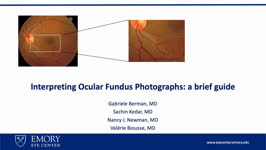 |
Interpreting Ocular Fundus Photographs: A Brief Guide | Gabriele Berman; Sachin Kedar; Nancy J. Newman; Valerie Biousse | This is a brief guide to the interpretation of the ocular fundus photograph. In this presentation we will describe the structures that comprise the normal ocular fundus followed the abnormalities that can be detected on fundus photographs. By the end of the presentation, learners should be able to d... | Fundus Photograph; Glaucoma; Papilledema; Retinal Detachment |
| 70 |
 |
Retinal Venous Occlusive Disease | Ali Alkhabbaz; James Brian Davis; Amanda Dean Henderson | Retinal venous occlusion (RVO) includes central retinal vein occlusion (CRVO), hemi-central retinal vein occlusion (HCRVO), and branch retinal vein occlusion (BRVO). The most important risk factor for RVO is hypertension, but other risk systemic factors include advanced age and cardiovascular diseas... | Branch Retinal Vein; BRVO; Central Retinal Vein; CRVO; HCRVO; Retinal Venous Occlusion |
| 71 |
 |
A Case of Orbital Meningioma Misdiagnosed as Non-arthritic Ischemic Optic Neuropathy | Hong Jiang, MD, PhD | A 62-year-old woman with hypertension, dyslipidemia, and mild cognitive impairment presented for a second opinion of her left eye visual loss for three months. She initially noticed a black spot in the lower visual field of her left eye and sought care from her local ophthalmologist. At that time, s... | Compressive Optic Neuropathy, Orbit MRI; Orbital Meningioma |
| 72 |
 |
Tacrolimus Optic Neuropathy | Hailey Mair, BS; Padmaja Sudhakar, MD | This PowerPoint slide deck will describe tacrolimus optic neuropathy which is a very rare form of optic neuropathy but remains a potential issue in many patients that receive this drug. | Optic Neuropathy; Tacrolimus; Tacrolimus Associated Visual Loss |
| 73 |
 |
Intracerebral Hemorrhage: Classification, Mechanisms, and Principles of Management | William Hills | Intracerebral hemorrhage accounts for approximately 10% of the 795,000 strokes that occur each year in the United States. Mortality is as high as 50% within 30 days of occurrence. There are several types of intracerebral hemorrhages based on inter-cranial location and mechanism. Early aggressive tre... | Epidural Hemorrhage; Intracranial Hemorrhage Management; Intraparenchymal Hemorrhage; Subarachnoid Hemorrhage; Subdural Hemorrhage |
| 74 |
 |
Retinal Artery Occlusive Disease | Ali Alkhabbaz, MD; James Brian Davis; Amanda Dean Henderson, MD | Retinal arterial occlusion includes ophthalmic artery occlusion (OAO), central retinal artery occlusion (CRAO), branch retinal arteriolar occlusion (BRAO), from proximal to distal. These can occur with or without retinal ischemia and may be permanent or transient. There are 4 subtypes of CRAO: non-a... | Branch Retinal Arteriolar Occlusion; Central Retinal Artery Occlusion; Giant Cell Arteritis; Ophthalmic Artery Occlusion |
| 75 |
 |
Raymond Cestan Syndrome | Srujay Pandiri; Sean Gratton | This is a brief narrated powerpoint that explains the clinical presentation of Raymond Cestan Syndrome. This is a rare brainstem stroke syndrome that presents with ipsilateral internuclear ophthalmoplegia and contralateral hemiparesis as well as other features. It is sometimes referred to as upper d... | Brainstem Stroke Syndromes; Internuclear Ophthalmoplegia; Pons |