Best known for his world-renowned neuro-ophthalmology unit based at the University of California, San Francisco, William Hoyt, MD collected here more than 850 of his best images covering a wide range of disorders.
William F. Hoyt, MD, Professor Emeritus of Ophthalmology, Neurology and Neurosurgery, Department of Ophthalmology, University of California, San Francisco.
NOVEL: https://novel.utah.edu/
TO
Filters: Collection: "ehsl_novel_wfh"
| Title | Description | Type | ||
|---|---|---|---|---|
| 826 |
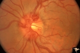 |
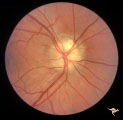
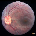
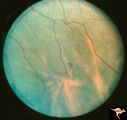
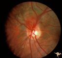
Venous Anomalies - Prepapillary Venous Convolutions (Congenital) | Prepapillary venous convolutions - congenital. 42 year old man. Incidental finding. Anatomy: Optic disc. Pathology: Prepapillary venous convolutions - congenital. Disease/Diagnosis: Prepapillary venous convolutions - congenital. Clinical: Asymptomatic. | Image |
| 827 |
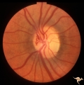 |
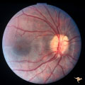
Venous Anomalies - Prepapillary Venous Convolutions (Congenital) | Prepapillary venous convolutions - congenital. Anatomy: Optic disc. Pathology: Prepapillary venous convolutions - congenital. Disease/Diagnosis: Prepapillary venous convolutions - congenital. Clinical: Asymptomatic. | Image |
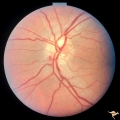
| 828 |
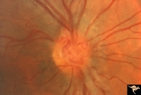 |
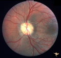
Venous Anomalies - Prepapillary Venous Convolutions (Congenital) | Prepapillary venous convolutions - congenital. Anatomy: Optic disc. Pathology: Prepapillary venous convolutions - congenital. Disease/Diagnosis: Prepapillary venous convolutions - congenital. Clinical: Asymptomatic. | Image |
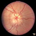
| 829 |
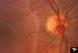 |
Venous Anomalies - Prepapillary Venous Convolutions (Congenital) | Prepapillary venous loop - congenital. Anatomy: Optic disc. Pathology: Prepapillary venous convolutions - congenital. Disease/Diagnosis: Prepapillary venous convolutions - congenital. Clinical: Asymptomatic. | Image |
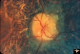
| 830 |
 |
Visible Drusen | PP21a: Right eye. Drusen barely visible. Note disc margin drusen at 1:00 and 2:30.; PP21b: Left eye shows multiple exposed drusen. Girl. Anatomy: Optic disc. Pathology: Drusen of the optic disc. Disease/Diagnosis: Drusen of the optic disc. Clinical: Normally functioning eye with drusen. | Image |
| 831 |
 |
Visible Drusen | PP24a. Right eye. Exposed drusen. There are inferior nerve fiber layer defects in the upper arcuate bundles. Optic disc is also hypoplastic. Anatomy: Optic disc. Pathology: Drusen of the optic disc. Disease/Diagnosis: Drusen of the optic disc. Clinical: Hypoplastic optic disc with drusen. | Image |
| 832 |
 |
Visible Drusen - Bilateral | PP22a: right eye. PP22b: Note bypass vein draining into the choroid at 8:00. Anatomy: Optic disc. Pathology: Drusen of the optic disc. Disease/Diagnosis: Drusen of the optic disc. Clinical: Normally functioning eye with drusen. | Image |
| 833 |
 |
Visible Drusen - Bilateral | PP22a: right eye with obvious exposed drusen. PP22 b: Note bypass vein draining into the choroid at 8:00. Anatomy: Optic disc. Pathology: Drusen of the optic disc. Disease/Diagnosis: Drusen of the optic disc. Clinical: Normally functioning eye with drusen. | Image |
| 834 |
 |
Visible Drusen with Retinitis Pigmentosa | Right eye. Optic disc drusen with retinitis pigmentosa. Note the marked narrowing of the retinal arterioles and the spectacular change in the peripapillary choroid. Anatomy: Optic disc. Pathology: Drusen of the optic disc. Disease/Diagnosis: Drusen of the optic disc. Clinical: Patient was nearly bli... | Image |
| 835 |
 |
Visible Drusen with Visual Field Loss | Right eye visual field combine with PP25a, b, & d. Anatomy: Optic disc. Pathology: Drusen of the optic disc. Disease/Diagnosis: Drusen of the optic disc. Clinical: Drusen disc with severe visual field defect. note the nasal visual field loss and the arcuate bundle defects. Central vision was 20/20. | Image |
| 836 |
 |
Visible Drusen with Visual Field Loss | Left eye visual field. Combine with PP25 a, b, & c. Anatomy: Optic disc. Pathology: Drusen of the optic disc. Disease/Diagnosis: Drusen of the optic disc. Clinical: Note marked constriction of visual field in all quadrants with central preservation of vision. | Image |
| 837 |
 |
Visible Drusen with Visual Field Loss | Left eye.16 year old girl: PP26b: buried drusen at the lower pole of the disc; PP26a: Visible drusen with visual field loss. Notice the thinning of the nerve fibers in both the superior and inferior arcuate bundles. PP26c: Goldmann visual field. Anatomy: Optic disc. Pathology: Drusen of the optic ... | Image |
| 838 |
 |
Visible Drusen with Visual Field Loss | Right eye.16 year old girl: PP26a: Visible drusen with visual field loss. Notice the thinning of the nerve fibers in both the superior and inferior arcuate bundles. PP26b: buried drusen; PP26c: Goldmann visual field. Anatomy: Optic disc. Pathology: Drusen of the optic disc. Disease/Diagnosis: Druse... | Image |
| 839 |
 |
Visible Drusen with Visual Field Loss | 16 year old girl: Drusen disc. Goldmann visual field. Anatomy: Optic disc. Pathology: Drusen of the optic disc. Disease/Diagnosis: Drusen of the optic disc. Clinical: Drusen disc with visual field loss. | Image |
| 840 |
 |
Visible Drusen with Visual Field Loss | PP25a: Left eye: Severe visual field defect. PP25b: right eye with exposed drusen and field loss: visual field defects; PP25c: right eye visual field PP25d: left eye visual field. Anatomy: Optic disc. Pathology: Drusen of the optic disc. Disease/Diagnosis: Drusen of the optic disc. Clinical: Dr... | Image |
| 841 |
 |
Visible Drusen with Visual Field Loss | PP25b right eye with drusen and severe visual field loss. Match with PP25a, c & d. Anatomy: Optic disc. Pathology: Drusen of the optic disc. Disease/Diagnosis: Drusen of the optic disc. Clinical: Drusen disc with servere visual field loss. | Image |
| 842 |
 |
Von Hippel Lindau Disease | Von Hippel Lindau Disease with a mini retinal tumor. Pair with R1_C4b. Anatomy: Retina. Pathology: Hemangioblastoma. Disease/Diagnosis: Von Hippel Lindau disease. Clinical: No visual symptoms. Imaging: Flourescien angiogram in R1_C4b. | Image |
| 843 |
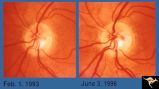 |
Von Hippel Lindau Disease | Von Hippel Lindau lesion on optic disc showing minimal increase in size over three year interval. Anatomy: Optic disc. Pathology: Hemangioblastoma. Disease/Diagnosis: Von Hippel Lindau disease. Clinical: Patient other eye was removed for hemangioblastoma. He has numerous hemangioblastomas of his spi... | Image |
| 844 |
 |
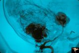
Von Hippel Lindau Disease | Von Hippel Lindau Disease with a mini retinal tumor. Flourescien angiogram shows how small tumor is. Pair with R1_C4a. Anatomy: Retina. Pathology: Hemangioblastoma. Disease/Diagnosis: Von Hippel Lindau disease. Clinical: No visual symptoms. Imaging: Flourescien angiogram. | Image |
| 845 |
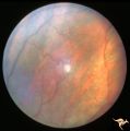 |
Von Hippel Lindau Disease | Von Hippel Lindau Disease. Retinal photograph showing small whitish hemangiomablastoma. Note the dilated arterial and venous channels entering and leaving the tumor. Anatomy: Retina. Pathology: Hemangioblastoma. Disease/Diagnosis: Von Hippel Lindau disease. Clinical: No visual symptoms. | Image |
| 846 |
 |
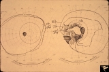
Von Hippel Lindau Disease (Hemangioblastoma of the Optic Disc) | Von Hippel Lindau Disease with a retinal hemangioblastoma on her optic disc. Anatomy: Optic disc. Pathology: Hemangioblastoma. Disease/Diagnosis: Von Hippel Lindau disease. Clinical: No visual symptoms. Patient had cerebellar ataxia. Imaging: R1_C1b is Arteriogram showing hemangioblastoma of the cer... | Image |
| 847 |
 |
Von Hippel Lindau Disease (Hemangioblastoma of the Optic Disc) | Von Hippel Lindau Disease; Arteriogram showing hemangioblastoma of the cerebellum and midbrain. Anatomy: Brain. Pathology: Hemangioblastoma. Disease/Diagnosis: Von Hippel Lindau disease. Clinical: No visual symptoms. Patient had cerebellar ataxia. Imaging: Arteriogram showing hemangioblastoma of the... | Image |
| 848 |
 |
Von Hippel Lindau Disease (Retinal Hemangioblastoma) | Von Hippel Lindau Disease with large peripheral retinal hemangioblastoma. View of the tumor. Larger artery entering and the vein leaving the tumor are evidence of rapid arteriovenous shunting within the tumor. Group with R1_C3b, R1_C3a, R1_C3d. Anatomy: Retina. Pathology: Hemangioblastoma. Disease/... | Image |
| 849 |
 |
Von Hippel Lindau Disease (Retinal Hemangioblastoma) | Von Hippel Lindau Disease with large retinal hemangioblastoma. Continued view of the arteriole and venous channels leading to the tumor. Group with R1_C3a, R1_C3c, R1_C3d. Anatomy: Retina. Pathology: Hemangioblastoma. Disease/Diagnosis: Von Hippel Lindau disease. Clinical: No visual symptoms. | Image |
| 850 |
 |
Von Hippel Lindau Disease (Retinal Hemangioblastoma) | Von Hippel Lindau Disease (Retinal Hemangioblastoma); Small hemangioblastoma on the disc margin at 10:00. Large peripheral hemangioblastoma out of view to the top right seen on R1_C2b. Anatomy: Optic disc. Pathology: Hemangioblastoma. Disease/Diagnosis: Von Hippel Lindau disease. Clinical: No visual... | Image |