Best known for his world-renowned neuro-ophthalmology unit based at the University of California, San Francisco, William Hoyt, MD collected here more than 850 of his best images covering a wide range of disorders.
William F. Hoyt, MD, Professor Emeritus of Ophthalmology, Neurology and Neurosurgery, Department of Ophthalmology, University of California, San Francisco.
NOVEL: https://novel.utah.edu/
TO
Filters: Collection: "ehsl_novel_wfh"
| Title | Description | Type | ||
|---|---|---|---|---|
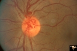
| 776 |
 |
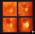
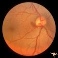
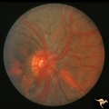
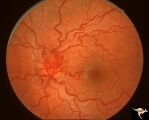
Vascular Complications of Drusen: Drusen Causing Loss of Superior Retinal Arterial Supply | PP32a: right; PP32b: left eye. Left eye has occlusion of superior branch of the central retinal artery at 11:30 with the inferior retinal artery supplying collateral to the superior retina. Notice the branch of the inferior retinal artery moves superiorly heading toward the upper retina. Drusen w... | Image |
| 777 |
 |
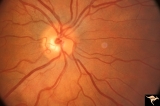
Vascular Disc Anomalies - Prepapillary Arterial Convolutions | Collection of prepapillary arterial congenital convolutions. Note: Typically involve the superior retinal arterioles. Purely arterial malformations (not arterial venous). Within the convolution in C, there are multiple tortuous loops. Not associated with cerebral vascular malformations and they do n... | Image |
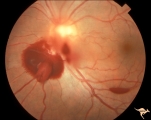
| 778 |
 |
Vascular Disc Anomalies - Prepapillary Arterial Convolutions | Hemorrhage from prepapillary arterial convolutions has resolved. Abnormal vessels which were the source of the bleeding. 30 year old man. 3.5 months following hemorrhage. Same patient as V_10. Anatomy: Optic disc. Pathology: Congenital prepapillary arterial convolutions with pre-retinal hemorrhage.... | Image |
| 779 |
 |
Vascular Disc Anomalies - Prepapillary Arterial Convolutions | Hemorrhage from prepapillary arterial convolutions. Note convolutions are inferior. 30 year old man. Same patient as V_11. Anatomy: Optic disc. Pathology: Congenital prepapillary arterial convolutions with pre-retinal hemorrhage. Disease/Diagnosis: Congenital arterial vascular anomaly. Clinical: Ac... | Image |
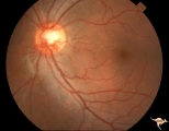
| 780 |
 |
Vascular Disc Anomalies - Prepapillary Arterial Convolutions | Prepapillary arterial convolutions. Left eye. Man. Anatomy: Optic disc. Pathology: Congenital prepapillary arterial convolutions. Disease/Diagnosis: Congenital arterial vascular anomaly. Clinical: Asymptomatic. | Image |
| 781 |
 |
Vascular Disc Anomalies - Prepapillary Arterial Convolutions | Prepapillary arterial convolutions. Incidental finding in patient being treated for acute myelogenous leukemia. Note hemorrhage at about 4:00 off the disc related to the leukemia. Arterial loops are not related to leukemia. Anatomy: Optic disc. Pathology: Congenital prepapillary arterial convolution... | Image |
| 782 |
 |
Vascular Disc Anomalies - Prepapillary Arterial Convolutions | Prepapillary arterial convolutions. 40 year old woman. Anatomy: Optic disc. Pathology: Congenital prepapillary arterial convolutions. Disease/Diagnosis: Congenital arterial vascular anomaly. Clinical: Asymptomatic. | Image |
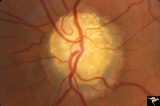
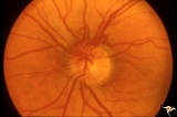
| 783 |
 |
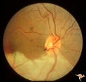
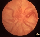
Vascular Disc Anomalies - Prepapillary Arterial Loop | Multiple prepapillary arterial loops involving superior and inferior retinal arterioles. Anatomy: Optic disc. Pathology: Congenital prepapillary arterial loop. Disease/Diagnosis: Congenital prepapillary arterial loop. Clinical: Asymptomatic. Patient presented with migraine. | Image |
| 784 |
 |
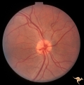
Vascular Disc Anomalies - Prepapillary Arterial Loop | Prepapillary arterial loop arising typically from the inferior retinal arterioles and projecting forward into the vitreous. 42 year old patient. Anatomy: Optic disc. Pathology: Congenital prepapillary arterial loop. Disease/Diagnosis: Congenital prepapillary arterial loop. Clinical: Asymptomatic. | Image |
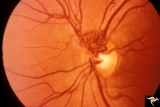
| 785 |
 |
Vascular Disc Anomalies - Prepapillary Arterial Loop | Small central prepapillary arterial loop. 30 year old woman. Anatomy: Optic disc. Pathology: Congenital prepapillary arterial loop. Disease/Diagnosis: Congenital prepapillary arterial loop. Clinical: Asymptomatic. | Image |
| 786 |
 |
Vascular Disc Anomalies - Prepapillary Arterial Loop | Complication of prepapillary arterial loop causing occlusion of the inferior retinal arterial and resulting inferior retinal infarction. Appears to be a black thrombus in the apex of the arterial loop. Anatomy: Optic disc. Pathology: Arterial loop with retinal artery infarction. Disease/Diagnosis: B... | Image |
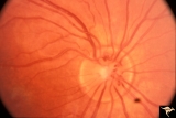
| 787 |
 |
Vascular Disc Anomalies - Prepapillary Arterial Loop | Prepapillary arterial loops. 36 year old woman. Anatomy: Optic disc. Pathology: Congenital prepapillary arterial loop. Disease/Diagnosis: Congenital prepapillary arterial loop. Clinical: Asymptomatic. | Image |
| 788 |
 |
Vascular Disc Anomalies - Retinal Arteriovenous Malformations | Wyburn-Mason Syndrome in man with orbital cranial component. After embolization of the orbital and cranial component, the retinal malformation involuted further. Retinal arteriovenous malformations. Post-treatment. Close up view of the disc. October 21, 1992. Same patient as V_23 and V_25. Anatomy: ... | Image |
| 789 |
 |
Vascular Disc Anomalies - Retinal Arteriovenous Malformations | Wyburn-Mason Syndrome in man with orbital cranial component. After embolization of the orbital and cranial component, the retinal malformation involuted further. Retinal arteriovenous malformations. Post-treatment. Wide angle view of slide V_24. October 21, 1992. Same patient as V_23 and V_24. Anat... | Image |
| 790 |
 |
Vascular Disc Anomalies - Retinal Arteriovenous Malformations | Wyburn-Mason Syndrome in man with orbital cranial component. Pre-treatment. Retinal arteriovenous malformations, pre-involution. April 27, 1992. Same patient as V_24 and V_25. Anatomy: Optic disc; Brain. Pathology: Arteriovenous malformation of retina and brain. Disease/Diagnosis: Wyburn-Mason syndr... | Image |
| 791 |
 |
Vascular Disc Anomalies - Retinal Arteriovenous Malformations | Retinal arteriovenous malformations. Wyburn-Mason Syndrome. Angiogram showing extension of vascular malformation up the right optic nerve (arrow) through the thalamus and into the right visual cortex. References #3 and #73. Anatomy: Brain. Pathology: Arteriovenous malformation. Disease/Diagnosis: Wy... | Image |
| 792 |
 |
Vascular Disc Anomalies - Retinal Arteriovenous Malformations | Retinal arteriovenous malformations. Partially involved. Same patient as V_27. Anatomy: Optic disc; Brain. Pathology: Arteriovenous malformation of retina and brain. Disease/Diagnosis: Wyburn-Mason syndrome. Clinical: Blindness in the involved eye, proptosis. | Image |
| 793 |
 |
Vascular Disc Anomalies - Retinal Arteriovenous Malformations | Retinal arteriovenous malformations. Anatomy: Optic disc. Pathology: Retinal arteriovenous malformation. Disease/Diagnosis: Wyburn-Mason syndrome. Clinical: Cerebral symptoms, questionable seizures. | Image |
| 794 |
 |
Vascular Disc Anomalies - Retinal Arteriovenous Malformations | Retinal arteriovenous malformations. Anatomy: Optic disc. Pathology: Retinal arteriovenous malformation. Disease/Diagnosis: Retinal arteriovenous malformation. Clinical: Reduced vision. | Image |
| 795 |
 |
Vascular Disc Anomalies - Retinal Arteriovenous Malformations | Retinal arteriovenous malformations. 11 year old. Anatomy: Optic disc. Pathology: Retinal arteriovenous malformation. Disease/Diagnosis: Retinal arteriovenous malformation. Clinical: Reduced visual function. | Image |
| 796 |
 |
Vascular Disc Anomalies - Retinal Arteriovenous Malformations | Retinal arteriovenous malformations of disc and adjacent retina. Anatomy: Optic disc. Pathology: Retinal arteriovenous malformation. Disease/Diagnosis: Retinal arteriovenous malformation. Clinical: Asymptomatic. | Image |
| 797 |
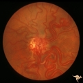 |
Vascular Disc Anomalies - Retinal Arteriovenous Malformations | Retinal arteriovenous malformations (Racemous angioma). Found in a 13 year old girl who had extension of this arteriovenous malformation up her right optic nerve into her thalamus and into her midbrain. Patient had large intra-cerebral AVM (Wyburn-Mason Syndrome). Patient died 10 years later of mass... | Image |
| 798 |
 |
Vascular Disc Anomalies - Retinal Arteriovenous Malformations | Retinal arteriovenous malformations with no known cerebral component. Anatomy: Optic disc. Pathology: Retinal arteriovenous malformation. Disease/Diagnosis: Retinal arteriovenous malformation. Clinical: Cerebral symptoms, questionable seizures. | Image |
| 799 |
 |
Vascular Disc Anomalies - Retinal Arteriovenous Malformations | Retinal arteriovenous malformations. Note ghost vessels, signs of involution within the malformation. Natural history is spontaneous involution of arterial loops within the malformation. Anatomy: Optic disc. Pathology: Retinal arteriovenous malformation. Disease/Diagnosis: Retinal arteriovenous malf... | Image |
| 800 |
 |
Vascular Disc Anomalies - Retinal Arteriovenous Malformations | Retinal arteriovenous malformations. Spontaneous involution. Bonnet-Dechaume-Blanc syndrome. Anatomy: Optic disc; Brain. Pathology: Arteriovenous malformation of retina and brain. Disease/Diagnosis: Wyburn-Mason syndrome. Clinical: Blindness in the involved eye. | Image |