Best known for his world-renowned neuro-ophthalmology unit based at the University of California, San Francisco, William Hoyt, MD collected here more than 850 of his best images covering a wide range of disorders.
William F. Hoyt, MD, Professor Emeritus of Ophthalmology, Neurology and Neurosurgery, Department of Ophthalmology, University of California, San Francisco.
NOVEL: https://novel.utah.edu/
TO
Filters: Collection: "ehsl_novel_wfh"
| Title | Description | Type | ||
|---|---|---|---|---|
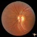
| 676 |
 |
Retinal Signs of Atheromatous Embolization | Retinal signs of atheromatous embolization. Arteriole wall changes produced by a stuttering atheromatous embolus. Note the beaded track of the cholesterol embolus in the inferior retinal arteriole. Anatomy: Retina. Pathology: Carotid atheromatous disease. Disease/Diagnosis: Carotid atheromatous vasc... | Image |
| 677 |
 |
Retinal Signs of Atheromatous Embolization | Retinal signs of atheromatous embolization in the superior temporal arteriole. Atheromatous emboli. Also note an embolus more distally in the inferior temporal arteriole. Left eye. Anatomy: Retina. Pathology: Carotid atheromatous disease. Disease/Diagnosis: Carotid atheromatous vascular disease. Cli... | Image |
| 678 |
 |
Retinal Signs of Atheromatous Embolization | Retinal signs of atheromatous embolization. Branch retinal artery occlusion from atheromatous debris. Note gray areas of retina including the macula indicating infarction. Anatomy: Retina. Pathology: Carotid atheromatous disease. Disease/Diagnosis: Carotid atheromatous vascular disease. Clinical: Su... | Image |
| 679 |
 |
Retinal Signs of Atheromatous Embolization | Retinal signs of atheromatous embolization. Documentation of atheromatous embolus appearing at bifurcation during photographic session. A shows no embolus and R3_A18b shows new embolus. Anatomy: Retina. Pathology: Carotid atheromatous disease. Disease/Diagnosis: Carotid atheromatous vascular disease... | Image |
| 680 |
 |
Retinal Signs of Atheromatous Embolization | Retinal signs of atheromatous embolization. Atheromatous emboli. Second view of inferior retinal arteriole with cholesterol embolus. Left eye. Pair with R3_A12a. Anatomy: Retina. Pathology: Carotid atheromatous disease. Disease/Diagnosis: Carotid atheromatous vascular disease. Clinical: Transient ri... | Image |
| 681 |
 |
Retinal Signs of Atheromatous Embolization | Retinal signs of atheromatous embolization. Central retinal artery occlusion at the level of the optic disc. The embolus that caused it can not be seen. Anatomy: Retina. Pathology: Carotid atheromatous disease. Disease/Diagnosis: Carotid atheromatous vascular disease. Clinical: Blindness. | Image |
| 682 |
 |
Retinal Signs of Atheromatous Embolization | Retinal signs of atheromatous embolization. Central retinal artery occlusion by soft atheromatous debris (mostly fibrin) causing blindness. Anatomy: Retina. Pathology: Carotid atheromatous disease. Disease/Diagnosis: Carotid atheromatous vascular disease. Clinical: Blindness. | Image |
| 683 |
 |
Retinal Signs of Atheromatous Embolization | Retinal signs of atheromatous embolization. Central retinal artery occlusion by soft atheromatous debris (mostly fibrin) causing blindness. All but one of the retinal arterioles have been converted to white strands. Anatomy: Retina. Pathology: Source of the embolic occlusion not determined. Disease/... | Image |
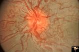
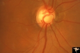
| 684 |
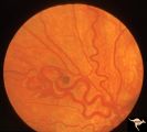 |
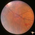
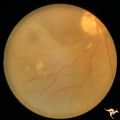
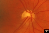
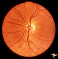
Retinocerebral Arteriovenous Malformation (Wyburn Mason Syndrome) | Retinocerebral arteriovenous malformation showing one major arteriovenous loop. (Cross reference with V12-28 this section). Cross reference with V12-28 this section, Anatomy: Optic disc. Pathology: Arteriovenous malformation. Disease/Diagnosis: Wyburn Mason Syndrome. Clinical: Single arteriovenous l... | Image |
| 685 |
 |
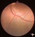
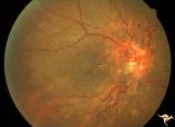
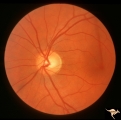
Retinocerebral Arteriovenous Malformation (Wyburn Mason Syndrome) | Retinocerebral arteriovenous malformation showing multiple arteriovenous shunts, both small and large. (Cross reference with V12-28 this section). Anatomy: Retina. Pathology: Arteriovenous malformation. Disease/Diagnosis: Wyburn Mason Syndrome. Clinical: Arteriovenous loop in the inferior temporal r... | Image |
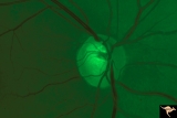
| 686 |
 |
Retinocerebral Arteriovenous Malformation (Wyburn Mason Syndrome) | Florid arteriovenous malformation of the optic disc and surrounding retina, Caput medusa (Cross reference with V12-28 this section). Anatomy: Retina. Pathology: Arteriovenous malformation. Disease/Diagnosis: Wyburn Mason Syndrome. | Image |
| 687 |
 |
Retinocerebral Arteriovenous Malformation (Wyburn Mason Syndrome) | Retinocerebral arteriovenous malformation with optic atrophy and central extension up the optic nerve into the brain. Ipsilateral facial involvement. Man from Thailand. Anatomy: Retina. Pathology: Arteriovenous malformation. Disease/Diagnosis: Wyburn Mason Syndrome. Clinical: Asian man with an exten... | Image |
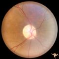
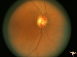
| 688 |
 |
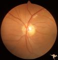
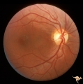
Segmental Atrophy - Altitudinal | Segmental optic atrophy - superior altitudinal. 55 year old man.1970. The cupping and the normal superior arteries are evidence against AION. Post ischemic, acquired. Anatomy: Optic disc. Pathology: Optic hemiatrophy. Disease/Diagnosis: Segmental atrophy - altitudinal. Clinical: Inferior visual fiel... | Image |
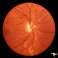
| 689 |
 |
Segmental Atrophy - Altitudinal | Segmental Optic Atrophy Superiorly - Altitudinal. Cause unknown. There is a cup. 1973. Anatomy: Optic disc. Pathology: Optic hemiatrophy. Disease/Diagnosis: Segmental atrophy - altitudinal. Clinical: Inferior visual field defect. | Image |
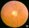
| 690 |
 |
Segmental Atrophy - Hemianopic (Band) Atrophy | Segmental Atrophy - Band atrophy in an eye with temporal hemianopia. Wyburn-Mason Syndrome extending to the chiasm. Left eye 1975. Anatomy: Optic disc. Pathology: Right sided chiasmal AVM. Disease/Diagnosis: Band atrophy due to chiasmal AVM and Wyburn-Mason Syndrome. Clinical: Blind right eye, temp... | Image |
| 691 |
 |
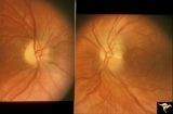
Segmental Atrophy - Hemianopic (Band) Atrophy | Segmental Atrophy - Band atrophy from right optic tract injury. Red free filter. Left eye. Has temporal hemianopia with band atrophy. Note loss of nasal nerve fiber layer. Old right optic tract injury. 1972. Pair with IIA2C_9a. Anatomy: Optic disc. Pathology: Right optic tract injury. Disease/Diagno... | Image |
| 692 |
 |
Segmental Atrophy - Hemianopic (Band) Atrophy | Segmental Atrophy - Band atrophy with horizontal cupping. Pituitary adenoma. Magnification of 14a. Pair with IIA2C_14a. 1975. Anatomy: Optic disc. Pathology: Chiasmal compression from pituitary adenoma in a cupped disc. Disease/Diagnosis: Band atrophy and cupping. Clinical: Temporal hemianopia. | Image |
| 693 |
 |
Segmental Atrophy - Hemianopic (Band) Atrophy | Segmental Atrophy - Band atrophy with horizontal cupping. Transverse cup. Pair with IIA2C_14b. 1975. Anatomy: Optic disc. Pathology: Chiasmal compression from pituitary adenoma in a cupped disc. Disease/Diagnosis: Band atrophy and cupping. Clinical: Temporal hemianopia. | Image |
| 694 |
 |
Segmental Atrophy - Hemianopic (Band) Atrophy | Segmental Atrophy - Band atrophy from right optic tract injury. Red free filter. Left eye. Has temporal hemianopia with band atrophy. Note loss of nasal nerve fiber layer. Old right optic tract injury. 1972. Pair with IIA2C_9b. Anatomy: Optic disc. Pathology: Right optic tract injury. Disease/Diagno... | Image |
| 695 |
 |
Segmental Atrophy - Hemianopic (Band) Atrophy | Segmental Atrophy - Band atrophy from right optic tract injury. This eye has a nasal hemianopia. Its disc shows temporal pallor with an intact nasal nerve fiber layer. Old right optic tract injury. 1986. Pair with IIA2C_8b. Anatomy: Optic disc. Pathology: Right optic tract injury. Disease/Diagnosis:... | Image |
| 696 |
 |
Segmental Atrophy - Hemianopic (Band) Atrophy | Segmental Atrophy - Band atrophy from right optic tract injury. Left eye. Has temporal hemianopia with band atrophy. Note loss of nasal nerve fiber layer. Old right optic tract injury. 1986. Pair with IIA2C_8a. Anatomy: Optic disc. Pathology: Right optic tract injury. Disease/Diagnosis: Homonymous h... | Image |
| 697 |
 |
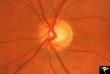
Segmental Atrophy - Hemianopic (Band) Atrophy | Segmental Atrophy - Band atrophy with papilledema. 1975. Patient had a right optic tract glioma. Anatomy: Optic disc. Pathology: Glioma of the right optic tract. Disease/Diagnosis: Twin peaks papilledema. Clinical: Left homonymous hemianopia. | Image |
| 698 |
 |
Segmental Atrophy - Hemianopic (Band) Atrophy | Segmental Atrophy - Magnification of IIA2C_02a. Band atrophy in an eye with temporal hemianopia. Wyburn-Mason Syndrome extending to the chiasm. Left eye. 1975. Right eye in patient was blind. Anatomy: Optic disc. Pathology: Right sided chiasmal AVM. Disease/Diagnosis: Band atrophy due to chiasmal A... | Image |
| 699 |
 |
Segmental Atrophy - Hemianopic (Band) Atrophy | Segmental Atrophy - Hemianopic (band) atrophy - Bilateral horizontal band atrophy secondary to old chiasmal trauma. Note the presence of arcuate nerve fibers and the absence of temporal and nasal nerve fibers. Note the sharp edged pallor of the nasal disc margin. Right eye. Pair with IIA2C_1b. 1985.... | Image |
| 700 |
 |
Segmental Atrophy - Hemianopic (Band) Atrophy | Segmental Atrophy - Hemianopic (band) atrophy - Bilateral horizontal band atrophy secondary to old chiasmal trauma. Note the presence of arcuate nerve fibers and the absence of temporal and nasal nerve fibers. Note the sharp edged pallor of the nasal disc margin. Right eye. Pair with IIA2C_1a. 1985.... | Image |