Collection of materials relating to neuro-ophthalmology as part of the Neuro-Ophthalmology Virtual Education Library.
NOVEL: https://novel.utah.edu/
TO
- NOVEL978
Filters: Collection: "ehsl_novel_novel"
| Title | Creator | Description | Subject | ||
|---|---|---|---|---|---|
| 401 |
 |
The Ventricles: Neuroanatomy Video Lab - Brain Dissections | Suzanne S. Stensaas, PhD | The ventricles are demonstrated and named on a model cast as well as in rotating 3D reconstructions. The production, function, circulation and removal of CSF produced by the choroid plexus is discussed using a diagram and then reviewed on frontal, axial and sagittal brain specimens and corresponding... | Ventricles; Brain; Dissection |
| 402 |
 |
Hypothalamus: Neuroanatomy Video Lab - Brain Dissections | Suzanne S. Stensaas, PhD | Gross specimens are used to demonstrate the area of the hypothalamus and its relationship to surrounding structures. Both endocrine and autonomic functions are explored using diagrams. Mention is made of the direct hypothalamic response to circulating hormones and other substances such as sodium. Th... | Hypothalamus; Brain; Dissection |
| 403 |
 |
Three Critical Vertical Pathways: Neuroanatomy Video Lab - Brain Dissections | Suzanne S. Stensaas, PhD | There is one motor and two sensory pathways that must be mastered. Pain and temperature from the body travel together and vibration and proprioception travel in another pathway each reaching perception in the cortex. Voluntary motor control starts in the cerebral cortex and connects with a motor neu... | Spinothalamic Tract; Dorsal Column-Medical Lemniscus Pathway; Posterior Column; Vertical Pathway; Brain; Dissection |
| 404 |
 |
The Spinal Cord & Monosynaptic Reflex: Neuroanatomy Video Lab - Brain Dissections | Suzanne S. Stensaas, PhD | The spinal cord's relationship to the foramina, discs and spinal nerves is demonstrated on a model. The dura, ganglia and rootlets are shown as well as the gray and white matter in gross sections at different levels. A model of the cord is used to demonstrate and describe the anatomy of a monosynapt... | Spinal Cord; Monosynaptic Reflex; Brain; Dissection |
| 405 |
 |
Limbic System: Neuroanatomy Video Lab - Brain Dissections | Suzanne S. Stensaas, PhD | The decision was made to present a simplified description of a much more complex system using animations to construct a 3D image. Papez circuit is shown on gross specimens with mention of its involvement in memory. The role of the amygdala in fear and the olfactory cortex in temporal lobe epilepsy a... | Limbic System; Hippocampus; Brain; Dissection |
| 406 |
 |
The Most Important Pathway: Motor Control: Neuroanatomy Video Lab - Brain Dissections | Suzanne S. Stensaas, PhD | The origin of the corticospinal tract in the cerebral cortex is traced through gross sections of the hemisphere and brain stem to the spinal cord. Using an animation, the terms upper and lower motor neuron are defined and clinical signs and symptom listed. | Corticospinal Tract; Cerebral Cortex; Motor Neuron; Brain; Dissection |
| 407 |
 |
The Unfixed Spinal Cord: Neuroanatomy Video Lab - Brain Dissections | Suzanne S. Stensaas, PhD | The spinal cord's relationship to the foramina, discs and spinal nerves is demonstrated on a model. The dura, ganglia and rootlets are shown as well as the gray and white matter in gross sections at different levels. A model of the cord is used to demonstrate and describe the anatomy of a monosynapt... | Unfixed Spinal Cord; Spinal Cord; Brain; Dissection |
| 408 |
 |
Olfactory System: Neuroanatomy Video Lab - Brain Dissections | Suzanne S. Stensaas, PhD | Beginning with the location of the sensory cells within the skull the axons are traced into the cranial cavity. Demonstration of the olfactory bulb, olfactory tract and it termination in the forebrain and temporal lobe are indicated. Trauma and meningiomas can produce loss of small (anosmia). Degene... | Olfactory System; Olfactory Bulb; Anosmia; Brain; Dissection |
| 409 |
 |
The Meninges: Neuroanatomy Video Lab - Brain Dissections | Suzanne S. Stensaas, PhD | The epidural, subdural and subarachnoid spaces are demonstrated and discussed with respect to trauma and disease. The relationship of the brainstem and cerebellum to the tentorium demonstrates the vulnerability of the brain stem to increased supratentorial pressure and herniation. Arachnoid granulat... | Meninges; Brain; Dissection |
| 410 |
 |
Orientation: The Planes of the Brain: Neuroanatomy Video Lab - Brain Dissections | Suzanne S. Stensaas, PhD | Terms such as anterior, posterior, inferior and superior are introduced with respect to the hemispheres as well as the brain stem. Terms such as rostral and caudal or dorsal and ventral can mean different things in different areas. Sections in three planes (frontal, axial, and sagittal) are demonstr... | Frontal; Axial; Sagittal; Brain; Dissection |
| 411 |
 |
The Visual Pathway: Neuroanatomy Video Lab - Brain Dissections | Suzanne S. Stensaas, PhD | A brief review of the anatomy of the eye and the photic stimulation of the receptors is followed by a gross exploration of the visual pathway from the optic nerve, chiasm, and tract to the thalamus stressing how the left part of the visual world reaches the right hemisphere. Visual fields are relate... | Visual Pathway; Brain; Dissections |
| 412 |
 |
The Normal Unfixed Brain: Neuroanatomy Video Lab - Brain Dissections | Suzanne S. Stensaas, PhD | The consistency and vulnerability of the brain is demonstrated along with the clear and glistening pia and arachnoid and the tough dura. The cushioning function of the CSF is stressed and the features are pointed out on the ventral surface. The uncus and temporal lobes are normal with arteries free ... | Brain; Dissections |
| 413 |
 |
Pituitary Tumor (Portuguese) | NANOS | Pituitary tumors are benign (non-cancerous) overgrowth of cells that make up the pituitary gland (the master gland that regulates other glands in the body). | Pituitary Tumor; Patient Brochure |
| 414 |
 |
Optic Disc Drusen (Portuguese) | NANOS | Optic disc drusen are abnormal deposits of protein-like material in the optic disc - the front part of the optic nerve. | Optic Disc Drusen; Patient Brochure |
| 415 |
 |
Optic Neuritis (Portuguese) | NANOS | In the most common form of optic neuritis, the optic nerve has been attacked by the body's overactive immune system and results in decreased vision. | Optic Neuritis; Patient Brochure |
| 416 |
 |
Transient Visual Loss (Portuguese) | NANOS | About transient visual loss. | Transient Visual Loss; Patient Brochure |
| 417 |
 |
Anterior Ischemic Optic Neuropathy (Portuguese) | NANOS | Loss of blood supply to the optic nerve results in diminished visual acuity. | Anterior Ischemic Optic Neuropathy; Patient Brochure |
| 418 |
 |
Photophobia (Portuguese) | NANOS | The symptoms of light sensitivity are: an uncomfortable sense of brightness, squinting, frequent blinking, and redness of the eye (especially if the eye is dry). Involuntary eye closure and excessive blinking is seen with blepharospasm. Individuals will tend to seclude themselves in darkness. | Photophobia; Patient Brochure |
| 419 |
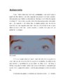 |
Anisocoria (Portuguese) | NANOS | Pupil in the right eye and left eye are not the same size. | Anisocoria; Patient Brochure |
| 420 |
 |
Myasthenia Gravis (Portuguese) | NANOS | This is an autoimmune condition where the body's immune system has damaged receptors on your muscles and can result in double vision or drooping lid. | Myesthenia Gravis; Patient Brochure |
| 421 |
 |
Progressive Supranuclear Palsy (Portuguese) | NANOS | About Progressive Supranuclear Palsy. | Progressive Supranuclear Palsy; Patient Brochure |
| 422 |
 |
Optic Nerve Sheath Meningioma (Portuguese) | NANOS | About optic nerve sheath meningioma. | Optic Nerve Sheath Meningioma; Patient Brochure |
| 423 |
 |
Blepharoespasm (Portuguese) | NANOS | Uncontrolled blinking, squeezing, and eyelid closure that occurs in both eyes without an apparent environmental cause. | Blepharospasm; Patient Brochure |
| 424 |
 |
Dry Eye Syndrome (Portuguese) | NANOS | People with abnormalities of the tear film are diagnosed with "dry eyes", but some patients with "dry eyes" may not feel that their eyes are "dry". Itching, burning, a scratchy sensation, a sensation that there is sand or grit in the eye, or intermittent blurring of the vision can all be symptoms of... | Dry Eye Syndrome; Patient Brochure |
| 425 |
 |
Homonymous Hemianopia (Portuguese) | NANOS | This refers to an absence of vision towards one side of the visual world in each eye. The damage that caused this problem is in the brain and not in the eyes. | Homonymous Hemianopia; Patient Brochure |