Best known for his world-renowned neuro-ophthalmology unit based at the University of California, San Francisco, William Hoyt, MD collected here more than 850 of his best images covering a wide range of disorders.
William F. Hoyt, MD, Professor Emeritus of Ophthalmology, Neurology and Neurosurgery, Department of Ophthalmology, University of California, San Francisco.
NOVEL: https://novel.utah.edu/
TO
1 - 25 of 11
| Title | Description | Type | ||
|---|---|---|---|---|
| 1 |
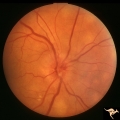 |
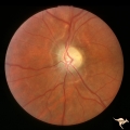
C109 Papillitis, Retrobulbar Neuritis | Optic papillitis after wasp sting. 57 year old woman. Right eye. Anatomy: Optic disc. Pathology: Axoplasmic stasis due to inflammation. Disease/ Diagnosis: Optic neuritis after wasp sting. Clinical: Visual loss after wasp sting. | Image |
| 2 |
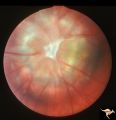 |
C17 Morning Glory Disc | "Morning Glory" disc. CT normal. Anatomy: Optic disc. Clinical: CT normal. | Image |
| 3 |
 |
C18 Morning Glory Disc | "Morning Glory" disc. 6 month old baby. Anatomy: Optic disc | Image |
| 4 |
 |
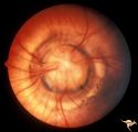
Congenitally Crowded Disc - Little Red Disc | Right eye: "little red disc". Congenitally blurred disc. 26 year old man. Anatomy: Optic disc Pathology: Normal variation of the optic disc Disease/Diagnosis: Normal variation of the optic disc. Congenital blurred disc. Little red disc. | Image |
| 5 |
 |
Post Papilledema, Secondary Optic Atrophy | Right eye. Post papilledema with chronic gliosis. arterial narrowing. ""high-water"" marks. Man. Anatomy: Optic disc. Pathology: Post papilledema. Disease/Diagnosis: Post papilledema with optic atrophy. | Image |
| 6 |
 |
Post Papilledema, Secondary Optic Atrophy | Left eye. Post papilledema with chronic gliosis. arterial narrowing. "high-water" marks. Man. Anatomy: Optic disc. Pathology: Post papilledema. Disease/Diagnosis: Post papilledema with optic atrophy. | Image |
| 7 |
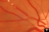 |
Retinal Signs of Atheromatous Embolization | Retinal signs of atheromatous embolization. Note shiny cholesterol plaques in retinal arterial. Anatomy: Retina. Pathology: Intraluminal cholesterol crystals. Disease/Diagnosis: Carotid atheromatous vascular disease. Clinical: No visual symptoms. | Image |
| 8 |
 |
Retinal Signs of Atheromatous Embolization | Retinal signs of atheromatous embolization. Atheromatous embolism in retinal arteriole branch with associated minimal opacification. Anatomy: Retina. Pathology: Carotid atheromatous disease. Disease/Diagnosis: Carotid atheromatous vascular disease. Clinical: Sudden inferior visual field loss. | Image |
| 9 |
 |
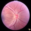
Unilateral Papilledema | Left eye. Has no optic cup. 24 year old obese woman. Anatomy: Optic disc. Pathology: Unilateral papilledema. Disease/Diagnosis: Idiopthatic intracranial hypertension, pseudotumor cerebri. Clinical: Woman, headache, transient visual obscurations. | Image |
| 10 |
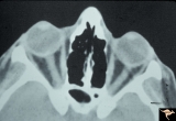 |
Unilateral Papilledema | CT scan showing equal thickening of the optic nerves. CT scan of no help in determining which eye has the papilledema. In this case, right eye has papilledema. Scan of patient depicted P_13a and P_13b. Anatomy: Optic disc. Pathology: Unilateral papilledema. Disease/Diagnosis: Idiopathic intracranial... | Image |
| 11 |
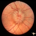 |
Unilateral Papilledema | Right eye. Has papilledema. Patient has pseudotumor cerebri. 24 year old obese woman. Anatomy: Optic disc. Pathology: Unilateral papilledema. Disease/Diagnosis: Idiopathic intracranial hypertension, pseudotumor cerebri. Clinical: Woman, headache, transient visual obscurations. | Image |
1 - 25 of 11