Best known for his world-renowned neuro-ophthalmology unit based at the University of California, San Francisco, William Hoyt, MD collected here more than 850 of his best images covering a wide range of disorders.
William F. Hoyt, MD, Professor Emeritus of Ophthalmology, Neurology and Neurosurgery, Department of Ophthalmology, University of California, San Francisco.
NOVEL: https://novel.utah.edu/
TO
1 - 25 of 9
| Title | Description | Type | ||
|---|---|---|---|---|
| 1 |
 |
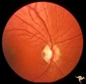
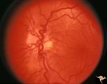
Buried Drusen | Young woman with pseudo papilledema from buried drusen with associated visual field defects. Barely visible in the upper arcuate nerve fibers is a slit like defect. Anatomy: Optic disc. Pathology: Drusen of the optic disc. Disease/Diagnosis: Drusen of the optic disc. Clinical notes: This patient had... | Image |
| 2 |
 |
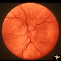
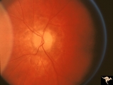
C06 Pits of the Optic Disc | Right eye. Temporal pit. 6 year old with see-saw nystagmus. Anatomy: Optic disc. Clinical: Six-year old with see-saw nystagmus. | Image |
| 3 |
 |
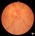
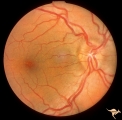
C103 Papillitis, Retrobulbar Neuritis | Optic neuritis in infectious mononucleosis. Anatomy: Optic disc. Pathology: Axoplasmic stasis due to inflammation. Disease/ Diagnosis: Optic neuritis with mononucleosis or Epstein Barr Virus. Clinical: Visual loss associated with mononucleosis. | Image |
| 4 |
 |
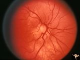
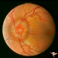
E08 Disc Swelling with Central Vein Occlusion | Pituitary adenoma with right chronic CRVO with optociliary bypass vessels. Anatomy: Optic disc; Retina. Pathology: Central retinal vein occlusion. Disease/ Diagnosis: Disc swelling due to central retinal vein occlusion. | Image |
| 5 |
 |
H27 Dysplasia with Hypoplasia (Elevated Dysplasia with Anomalous Vessels) | Right eye. Elevated hypoplastic dysplasia with anomalous vessels. Same patient as H_28. Anatomy: Optic disc. Pathology: Dysplasia of the optic disc. Disease/ Diagnosis: Elevated dysplasia with hypoplasia. | Image |
| 6 |
 |
H28 Dysplasia with Hypoplasia (Elevated Dysplasia with Anomalous Vessels) | Left eye. Elevated hypoplastic dysplasia with tortuous anomalous vessels. Same patient as H_27. Anatomy: Optic disc. Pathology: Dyplasia of the optic disc. Disease/ Diagnosis: Elevated dysplasia with hypoplasia. | Image |
| 7 |
 |
Resolution of Papilledema Following Optic Nerve Sheath Decompression (ONSD) | Left eye. 17 year old boy. Cryptococcal meningitis. Resolution of papilledema following optic nerve sheath decompression (ONSD) on November 1, 1974. Same eye as P_53a in January 1975. Atrophic, resolved disc. Note "high-water" marks. Visual acuity was 20/40. Anatomy: Optic disc. Pathology: Papilled... | Image |
| 8 |
 |
Unilateral Papilledema | Unilateral papilledema in Pseudotumor cerebri. Right eye. Has no cup. Woman. Anatomy: Optic disc. Pathology: Unilateral papilledema. Disease/Diagnosis: Idiopathic intracranial hypertension, pseudotumor cerebri. Clinical: Woman, headache. | Image |
| 9 |
 |
Unilateral Papilledema | Left eye. Has chronic papilledema. Woman. Anatomy: Optic disc. Pathology: Unilateral papilledema. Disease/Diagnosis: Idiopathic intracranial hypertension, pseudotumor cerebri. Clinical: Woman, headache. | Image |
1 - 25 of 9