Best known for his world-renowned neuro-ophthalmology unit based at the University of California, San Francisco, William Hoyt, MD collected here more than 850 of his best images covering a wide range of disorders.
William F. Hoyt, MD, Professor Emeritus of Ophthalmology, Neurology and Neurosurgery, Department of Ophthalmology, University of California, San Francisco.
NOVEL: https://novel.utah.edu/
TO
Filters: Date: "1973" Collection: "ehsl_novel_wfh"
1 - 25 of 18
| Title | Description | Type | ||
|---|---|---|---|---|
| 1 |
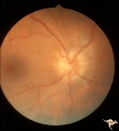 |
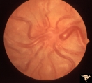
Bilateral Papilledema | Left eye. Chronic Bilateral Papilledema. Anatomy: Optic disc. Pathology: Chronic bilateral papilledema. Disease/Diagnosis: Pseudotumor long standing. Clinical notes: Chronic headache; Obesity. | Image |
| 2 |
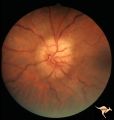 |
Bilateral Papilledema | Chronic Bilateral Papilledema. Anatomy: Optic disc. Pathology: Chronic bilateral papilledema. Disease/Diagnosis: Pseudotumor long standing. Clinical notes: Chronic headache; Obesity. | Image |
| 3 |
 |
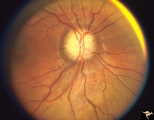
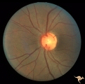
Bilateral Papilledema with Cyanotic Heart Disease | Bilateral Papilledema with cyanotic heart disease in a young boy. Anatomy: Optic disc. Pathology: Papilledema. Disease/Diagnosis: Pseudotumor due to cyanotic heart disease. Clinical notes: Young boy with clubbing. | Image |
| 4 |
 |
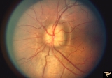
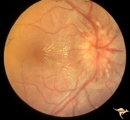
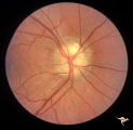
Bilateral Papilledema with Exudative Retinopathy | Bilateral Papilledema with exudative retinopathy from vitamin A toxicity. Young boy. Near blind. Anatomy: Optic disc; Retina. Pathology: Bilateral papilledema; exudative retinopathy. Disease/Diagnosis: Hypervitaminosis A causing blindness. Clinical notes: Nearly blind; Headache. | Image |
| 5 |
 |
Buried and Visible Drusen | PP_19b: right eye : visible drusen in an eleven year old girl; PP_19a: left eye with buried drusen. Anatomy: Optic disc Pathology: Drusen of the optic disc Disease/Diagnosis: Drusen of the optic disc Clinical: Normally functioning eye with drusen. | Image |
| 6 |
 |
Buried and Visible Drusen | PP_19a Left eye with buried drusen. PP_19b: right eye : visible drusen. Eleven year old girl. Anatomy: Optic disc. Pathology: Drusen of the optic disc. Disease/Diagnosis: Drusen of the optic disc. Clinical notes: Normally functioning eye with drusen. | Image |
| 7 |
 |
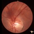
C202 Papillitis with Macular Star Cat Scratch Disease. | Proven Bartonella neuroretinitis. 23 year old man. Ocular disc edema with macular star (ODEMS). Anatomy: Optic disc; Retina. Pathology: Axoplasmic stasis due to inflammation; Exudate in Henle's layer. Neuroretinitis due to Bartonella Henslae (or cat scratch). Clinical: Visual blurring; Optic disc sw... | Image |
| 8 |
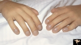 |
Cyanotic Heart Disease with Clubbing of Fingernails | Note the cyanotic nail beds and clubbing. Anatomy: Optic disc. Pathology: Papilledema. Disease/Diagnosis: Pseudotumor due to cyanotic heart disease. Clinical: Young boy with clubbing. | Image |
| 9 |
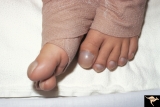 |
Cyanotic Heart Disease with Clubbing of Toes | Bilateral Papilledema with cyanotic heart disease. Anatomy: Optic disc. Pathology: Papilledema. Disease/Diagnosis: Pseudotumor due to cyanotic heart disease. Clinical: Young boy with clubbing. | Image |
| 10 |
 |
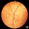
E07 Disc Swelling with Central Vein Occlusion | 24 year old male. Papillophlebitis (CRVO) with optic disc edema. Right eye. Anatomy: Optic disc; Retina. Pathology: Central retinal vein occlusion. Disease/ Diagnosis: Disc swelling due to central retinal vein occlusion. Clinical: ??Branch retinal artery occlusion [sic]. | Image |
| 11 |
 |
H13 Panhypoplasia | Right eye. Blind baby. Severe hypoplasia with blond fundus. Same patient as H_14. Anatomy: Optic disc. Pathology: Hypoplasia of the optic nerve. Disease/ Diagnosis: Hypoplasia. Imaging: Hypoplasia of the optic nerve. | Image |
| 12 |
 |
H14 Panhypoplasia | Left eye. Blind baby. Severe hypoplasia with blond fundus. Same patient as H_13. Anatomy: Optic disc. Pathology: Hypoplasia of the optic nerve. Disease/ Diagnosis: Hypoplasia. | Image |
| 13 |
 |
Late Complications of Drusen | PP33a: right disc shows pallor and small calcified crystals on the disc surface. PP33: left disc shows calcified specs on temporal sector of the disc. Florid drusen in young patients changes over time to assume this appearance. Anatomy: Optic disc. Pathology: Drusen of the optic disc. Disease/Dia... | Image |
| 14 |
 |
Late Complications of Drusen | PP33a: right disc shows pallor and small calcified crystals on the disc surface. PP33b: left disc shows calcified specs on temporal sector of the disc. Florid drusen in young patients changes over time to assume this appearance. Anatomy: Optic disc. Pathology: Drusen of the optic disc. Disease/Di... | Image |
| 15 |
 |
Vascular Disc Anomalies - Retinal Arteriovenous Malformations | Retinal arteriovenous malformations. Anatomy: Optic disc. Pathology: Retinal arteriovenous malformation. Disease/Diagnosis: Retinal arteriovenous malformation. Clinical: Reduced vision. | Image |
| 16 |
 |
Venous Anomalies - Exit Anomalies | Disc edge veins of Kraupa. 35 year old woman. Note that the arterial branches all appear to be cilioretinal. Empty disc. Anatomy: Optic disc. Pathology: Congenital anomaly, exit anomaly. Disease/Diagnosis: Exit anomaly, edge veins. Clinical: Asymptomatic. | Image |
| 17 |
 |
Visible Drusen | PP24a. Right eye. Exposed drusen. There are inferior nerve fiber layer defects in the upper arcuate bundles. Optic disc is also hypoplastic. Anatomy: Optic disc. Pathology: Drusen of the optic disc. Disease/Diagnosis: Drusen of the optic disc. Clinical: Hypoplastic optic disc with drusen. | Image |
| 18 |
 |
Von Hippel Lindau Disease Associated with Increased ICP | Von Hippel Lindau Disease; Optic disc lesion with hemorrhage from it in a patient with acute intracranial pressure elevation from a posterior fossa hemangioblastoma. Anatomy: Optic disc. Pathology: Hemangioblastoma. Disease/Diagnosis: Von Hippel Lindau disease. Clinical: Headache. | Image |
1 - 25 of 18