Best known for his world-renowned neuro-ophthalmology unit based at the University of California, San Francisco, William Hoyt, MD collected here more than 850 of his best images covering a wide range of disorders.
William F. Hoyt, MD, Professor Emeritus of Ophthalmology, Neurology and Neurosurgery, Department of Ophthalmology, University of California, San Francisco.
NOVEL: https://novel.utah.edu/
TO
Filters: Date: "1971" Collection: "ehsl_novel_wfh"
| Title | Description | Type | ||
|---|---|---|---|---|
| 1 |
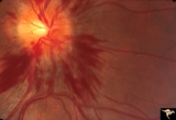 |
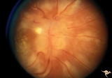
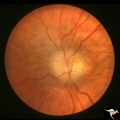
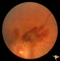
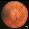
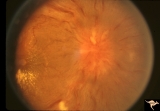
Bilateral Hemorrhagic Papilledema from Saggital Sinus Thrombosis | Left eye. 20 year old woman on oral contraceptives. Bilateral hemorrhagic Papilledema from sagittal sinus thrombosis. Anatomy: Optic disc. Pathology: Papilledema; hemorrhagic papilledema. Disease/Diagnosis: Superior saggital sinus thrombosis due to BCP use. Clinical notes: Chronic headache. | Image |
| 2 |
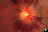 |
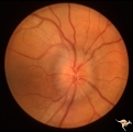
Bilateral Hemorrhagic Papilledema from Saggital Sinus Thrombosis | Right eye. 20 year old woman on oral contraceptives. Bilateral hemorrhagic Papilledema from sagittal sinus thrombosis. Anatomy: Optic disc. Pathology: Papilledema; hemorrhagic papilledema. Disease/Diagnosis: Superior sagittal sinus thrombosis due to BCP use. Clinical notes: Chronic headache. | Image |
| 3 |
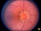 |
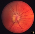
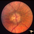
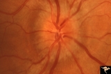
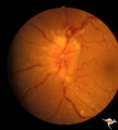
Bilateral Papilledema | Right eye. Bilateral Papilledema in patient with cardiopulmonary insufficiency. Woman. Anatomy: Optic disc. Pathology: Papilledema. Disease/Diagnosis: cardiopulmonary insufficiency causing intracranial hypertension. Clinical notes: headache. | Image |
| 4 |
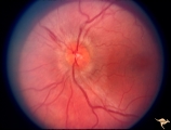 |
Bilateral Papilledema | Left eye. Bilateral Papilledema in patient with cardiopulmonary insufficiency. Woman. Anatomy: Optic disc. Pathology: Papilledema. Disease/Diagnosis: Cardiopulmonary insufficiency causing intracranial hypertension. Clinical notes: Headache. | Image |
| 5 |
 |
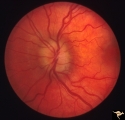
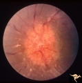
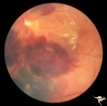
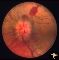
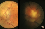
Bilateral Severe Hemorrhagic Papilledema | Right eye. Bilateral hyperacute papilledema with rapid blindness associated with dural sinus occlusion. Both eyes were nearly blind. Young man. Anatomy: Optic disc. Pathology: Papilledema. Disease/Diagnosis: Bilateral hyperacute papilledema | Image |
| 6 |
 |
Bilateral Severe Hemorrhagic Papilledema | Left eye. Bilateral hyperacute papilledema with rapid blindess associated with dural sinus occlusion. Both eyes were nearly blind. Boy. | Image |
| 7 |
 |
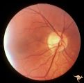
Buried Drusen | Buried drusen; PP_13a: Right eye. Note lumpy disc margin, especially temporally. Also note absence of optic cup. Excellent example of pseudo papilledema with buried drusen. Anatomy: Optic disc. Pathology: Drusen of the optic disc. Disease/Diagnosis: Drusen of the optic disc. Clinical notes: Patient ... | Image |
| 8 |
 |
Buried Drusen | Buried drusen. Left eye. Note lumpy disc margin, especially temporally. Also note absence of optic cup. Excellent example of pseudo papilledema with buried drusen. Pair with PP_13a. Anatomy: Optic disc. Pathology: Drusen of the optic disc. Disease/Diagnosis: Drusen of the optic disc. Clinical notes... | Image |
| 9 |
 |
C38 Anomalous Pale Disc | Megalopapilla in -8 myopic eye. Right eye. Anatomy: Optic disc. Clinical: High myope. | Image |
| 10 |
 |
Chronic Atrophic Papilledema | Left eye. Left eye blind. Chronic Atrophic Papilledema. Obese woman (300 lbs) with large tentorial meningioma. "Pseudo Pseudotumor". Anatomy: Optic disc. Pathology: Papilledema. Disease/Diagnosis: Papilledema from large tentorial meningioma. | Image |
| 11 |
 |
Chronic Atrophic Papilledema | Right eye. Chronic Atrophic Papilledema. Obese woman (300 lbs) with large tentorial meningioma. "Pseudo Pseudotumor" Anatomy: Optic disc. Pathology: Papilledema. Disease/Diagnosis: Papilledema from large tentorial meningioma. | Image |
| 12 |
 |
Chronic Papilledema in Resolution. Sequence | Left eye at presentation. Chronic papilledema. Anatomy: Optic disc Pathology: Papilledema Disease/Diagnosis: Papilledema | Image |
| 13 |
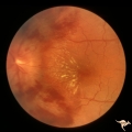 |
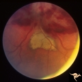
Chronic Papilledema with Hemorrhagic and Exudative Complications | Left eye one month after presentation. Resolving hemorrhage. Chronic papilledema with hemorrhagic and exudative complications due to Pseudotumor cerebri. Anatomy: Optic disc. Pathology: Papilledema. Disease/Diagnosis: Chronic papilledema with hemorrhagic and exudative complications | Image |
| 14 |
 |
Chronic Papilledema with Hemorrhagic and Exudative Complications | Left eye at presesntation. Chronic papilledema with hemorrhagic and exudative complications due to Pseudotumor cerebri. Anatomy: Optic disc Pathology: Papilledema Disease/Diagnosis: Chronic papilledema with hemorrhagic and exudative complications. | Image |
| 15 |
 |
Chronic Papilledema with Hemorrhagic and Exudative Complications | Left eye one month after presentation. View below of resolving subretinal hemorrhage. Chronic papilledema with hemorrhagic and exudative complications due to Pseudotumor cerebri. | Image |
| 16 |
 |
Chronic Papilledema with Hemorrhagic and Exudative Complications | Left eye one month after presentation. View above of resolving preretinal hemorrhage. Chronic papilledema with hemorrhagic and exudative complications due to Pseudotumor cerebri. | Image |
| 17 |
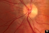 |
Crowded Disc | PP7a: right eye crowded disc with blurred margin. Note anomalous vascular pattern; PP7b- left disc is cupless disc and normal. 10 year old girl with gonadal dysgenesis and growth retardation. Anatomy: Optic disc Pathology: Normal variation of the optic disc Disease/Diagnosis: Normal variation of the... | Image |
| 18 |
 |
D101 Disc Edema with Systemic Lupus | Unilateral disc swelling with narrowed arterioles. No decrease in visual acuity or field. 19 year old woman. Patient died of cerebral lupus within two months. Optociliary veins dumping into disc edge at 4:00, 9:00, and 11:00. Anatomy: Optic disc. Pathology: Axoplasmic stasis due to vasculitis (Lupu... | Image |
| 19 |
 |
D102 Disc Edema with Systemic Lupus | 28 year old woman. Vision 20/20 but blind spot enlarged. Same patient as D1_03. Right eye. Anatomy: Optic disc. Pathology: Axoplasmic stasis due to vasculitis (Lupus). Disease/ Diagnosis: Lupus papillopathy. Clinical: Normal vision with enlarged blind spot on visual field. | Image |
| 20 |
 |
D103 Disc Edema with Systemic Lupus | 28 year old woman with systemic Lupus erythematosus. Vision 20/20 but blind spot enlarged. Same patient as D1_02. Magnified. Anatomy: Optic disc. Pathology: Axoplasmic stasis due to vasculitis (Lupus). Disease/ Diagnosis: Lupus papillopathy. Clinical: Normal vision with enlarged blind spot on visual... | Image |
| 21 |
 |
Early Papilledema due to Brain Tumor - Resolving | Left eye. Same eye as P_34a. One month post op, papilledema resolving. Boy. Anatomy: Optic disc. Pathology: Papilledema. Disease/Diagnosis: Papilledema from posterior fossa hemangioblastoma. | Image |
| 22 |
 |
Early Papilledema due to Tumor | Left eye. Asymmetric Papilledema with posterior fossa hemangioblastoma. Left - moderate papilledema. Blurring of disc. Young man. Anatomy: Optic disc. Pathology: Papilledema. Disease/Diagnosis: Papilledema from posterior fossa hemangioblastoma. | Image |
| 23 |
 |
F205 Optic Nerve Sheath Meningioma | Optic nerve meningioma of right optic nerve. Progressive visual field loss. Notice macular star and "cotton wool" spots. Anatomy: Optic disc. Pathology: Chronic optic disc swelling caused by optic nerve sheath meningioma. Disease/ Diagnosis: Chronic optic disc swelling caused by optic nerve sheath m... | Image |
| 24 |
 |
F206 Intracavernous Meningioma Extending Into the Orbit | Intracavernous meningioma extending into the orbit. Female patient. Anatomy: Optic disc. Pathology: Intracavernous meningioma. Disease/ Diagnosis: Neoplastic papillopathy. | Image |
| 25 |
 |
F2b09 Optic Disc Swelling from Malignant Optic Nerve Glioma | Malignant optic nerve glioma of adulthood with blindness and optic disc edema. Right image shows white material extruded from the swollen optic disc. This material is myelin being squeezed into the eye from the nerve infarction. Autopsy specimen of this eye shown in F2b_10. Reference: Hoyt WF, Meshe... | Image |