Best known for his world-renowned neuro-ophthalmology unit based at the University of California, San Francisco, William Hoyt, MD collected here more than 850 of his best images covering a wide range of disorders.
William F. Hoyt, MD, Professor Emeritus of Ophthalmology, Neurology and Neurosurgery, Department of Ophthalmology, University of California, San Francisco.
NOVEL: https://novel.utah.edu/
TO
Filters: Date: "1988" Collection: "ehsl_novel_wfh"
1 - 25 of 15
| Title | Description | Type | ||
|---|---|---|---|---|
| 1 |
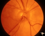 |
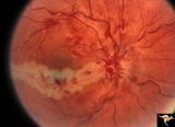
A402a Disc Swelling, Vitreous Effects | Posterior vitreous detachment with vitreo papillary adherence to the optic disc. See fluorescein angiogram A43b. Anatomy: Optic disc; Vitreous. Pathology: Posterior vitreous detachment with vitreo papillary adherence to the optic disc. Disease/ Diagnosis: Disc swelling due to vitreo papilary adheren... | Image |
| 2 |
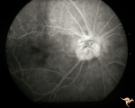 |
A403b Disc Swelling, Vitreous Effects | Fluorescein angiogram shows fluorescein leaking around entire disc where attachment of vitreous exists. Refers to A402a. Anatomy: Optic disc; Vitreous. Pathology: Disc swelling due to posterior vitreal detachment. Disease/ Diagnosis: Disc swelling due to posterior vitreal detachment. Clinical: float... | Image |
| 3 |
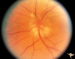 |
C111 Papillitis, Retrobulbar Neuritis | AIDS papillitis. Male. Anatomy: Optic disc. Pathology: Axoplasmic stasis due to inflammation. Disease/ Diagnosis: AIDS papillitis. Clinical: Visual symptoms. | Image |
| 4 |
 |
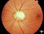
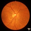
C27 Empty Disc | Multiple cilioretinal arteries. Chronic interstitial nephritis. Renal and optic disc dysplasia. Papilorenal Syndrome (PRS). No central retinal artery. Anatomy: Optic disc. | Image |
| 5 |
 |
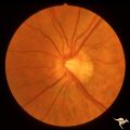
C28 Empty Disc | Left eye. Woman. Multiple cilioretinal vessels. Visual function normal. Anatomy: Optic disc. | Image |
| 6 |
 |
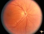
E10 Disc Swelling with Central Vein Occlusion | Cilioretinal artery infarction after a central retinal vein occlusion. Anatomy: Optic disc; Retina. Pathology: Central retinal vein occlusion. Disease/ Diagnosis: Disc swelling due to central retinal vein occlusion. | Image |
| 7 |
 |
H44 Segmental Hypoplasia, Retinal, Nasal Hypoplasia | Bilateral nasal hypoplasia with bilateral flag-like temporal field defect. Right eye. Same patient as H_45. Anatomy: Optic disc. Pathology: Nasal segmental disc hypoplasia. Disease/ Diagnosis: Congenital anomaly. | Image |
| 8 |
 |
H45 Segmental Hypoplasia, Retinal-Nasal Hypoplasia | Bilateral nasal hypoplasia with bilateral flag-like temporal field defect. Left eye. Same patient as H_44. Anatomy: Optic disc. Pathology: Nasal segmental disc hypoplasia. Disease/ Diagnosis: Congenital anomaly. | Image |
| 9 |
 |
H57 Superior Segmental Optic Hypoplasia (SSOH) Topless Disc Syndrome | Note, in addition to SSOH, generalized hypoplasia of nerve. Anatomy: Optic disc. Pathology: Superior segmental optic hypoplasia (SSOH). Disease/ Diagnosis: Congenital anomaly. | Image |
| 10 |
 |
H64 Superior Segmental Optic Hypoplasia (SSOH) Topless Disc Syndrome | Bilateral SSOH. Same patient as H_65 and H_66. Anatomy: Optic disc. Pathology: Superior segmental optic hypoplasia (SSOH). Disease/ Diagnosis: Congenital anomaly. | Image |
| 11 |
 |
H65 Superior Segmental Optic Hypoplasia (SSOH) Topless Disc Syndrome | Bilateral SSOH. Same patient as H_64 and H_66. Anatomy: Optic disc. Pathology: Superior segmental optic hypoplasia (SSOH). Disease/ Diagnosis: Congenital anomaly. | Image |
| 12 |
 |
H66 Superior Segmental Optic Hypoplasia (SSOH) Topless Disc Syndrome | Visual field. Same patient as H_65 and H_64. Anatomy: Optic disc. Pathology: Superior segmental optic hypoplasia (SSOH). Disease/ Diagnosis: Congenital anomaly. | Image |
| 13 |
 |
H75 Superior Segmental Optic Hypoplasia (SSOH) Topless Disc Syndrome | SSOH with hypoplasia of whole disc. Right eye. Anatomy: Optic disc. Pathology: Superior segmental optic hypoplasia (SSOH). Disease/ Diagnosis: Congenital anomaly. | Image |
| 14 |
 |
H91 Occipital Hemianoptic Hypoplasia | Left eye with temporal field defect shows trans-synaptic band atrophy. Same patient as H_92. Anatomy: Optic disc. Pathology: Occipital hemianoptic hypoplasia. Disease/ Diagnosis: Congenital defect of the occipital lobe. | Image |
| 15 |
 |
H92 Occipital Hemianoptic Hypoplasia | Right eye. Same patient as H_91. Anatomy: Optic disc. Pathology: Occipital hemianoptic hypoplasia. Disease/ Diagnosis: Congenital defect of the occipital lobe. | Image |
1 - 25 of 15