Best known for his world-renowned neuro-ophthalmology unit based at the University of California, San Francisco, William Hoyt, MD collected here more than 850 of his best images covering a wide range of disorders.
William F. Hoyt, MD, Professor Emeritus of Ophthalmology, Neurology and Neurosurgery, Department of Ophthalmology, University of California, San Francisco.
NOVEL: https://novel.utah.edu/
TO
Filters: Date: "1976" Collection: "ehsl_novel_wfh"
1 - 25 of 14
| Title | Description | Type | ||
|---|---|---|---|---|
| 1 |
 |
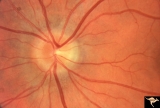
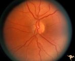
Buried Drusen | Excellent example of pseudo papilledema with sub surface drusen at 10:00 and 1:00. Anatomy: Optic disc. Pathology: Drusen of the optic disc. Disease/Diagnosis: Drusen of the optic disc. Clinical notes: Normally functioning eye with drusen. | Image |
| 2 |
 |
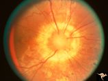
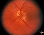
C115 Papillitis, Retrobulbar Neuritis | Demyelinative optic neuropathy with mild disc swelling. This eye had a large central scotoma. Note the bland disc margin swelling from 2:00 to 4:00. This swelling constitutes spill over edema from the main focus of the neuritis which lies behind the eyeball. Visual acuity was 2200. Anatomy: Optic di... | Image |
| 3 |
 |
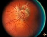
C19 Morning Glory Disc | Bilateral "Morning Glory" disc. Right eye. Man. Pair with C_20. Anatomy: Optic disc. | Image |
| 4 |
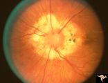 |
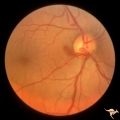
C20 Morning Glory Disc | Bilateral "Morning Glory" disc. Left eye. Man. Pair with C_19. Anatomy: Optic disc. | Image |
| 5 |
 |
H10 Panhypoplasia | Cruzon's Disease. 26 year old man. Right eye. Mild hypoplasia. Son of patient in H_11 and H_12. Same patient in H_31. Father of patient in H_32. Anatomy: Optic disc. Pathology: Hypoplasia of the optic nerve. Disease/ Diagnosis: Hypoplasia. | Image |
| 6 |
 |
H11 Panhypoplasia | Cruzon's Disease. 47 year old woman. Right eye. Mild hypoplasia. Mother of patient in H_10 and H_31. Same patient as H_12. Grandmother of patient in H_32. Anatomy: Optic disc. Pathology: Hypoplasia of the optic nerve. Disease/ Diagnosis: Hypoplasia. | Image |
| 7 |
 |
H12 Panhypoplasia | Cruzon's Disease. 47 year old woman. Left eye. Mild hypoplasia. Mother of patient in H_10 and H_31. Same patient as H_11. Grandmother of patient in H_32. Anatomy: Optic disc. Pathology: Hypoplasia of the optic nerve. Pathology: Hypoplasia of the optic nerve. Disease/ Diagnosis: Hypoplasia. | Image |
| 8 |
 |
H31 Dysplasia with Hypoplasia (Elevated Hysplasia with Anomalous Vessels) | Left eye. 26 year old man. Dysplasia with hypoplasia. Father of patient in H_32. Same patient as H_10. Son of patient in H_11 an H_12. Anatomy: Optic disc. Pathology: Dysplasia of the optic disc. Disease/ Diagnosis: Elevated dysplasia with hypoplasia. | Image |
| 9 |
 |
H32 Dysplasia with Hypoplasia (Elevated Dysplasia with Anomalous Vessels) | Left eye. 6 year old boy. Severe dysplasia. Elevated dysplasia with medullated (myelinated) nerve fibers and anomalous vessels. Son of patient in H_31 and H_10. Grandson of patient in H_11 an H_12. Anatomy: Optic disc. Pathology: Dysplasia of the optic disc. Disease/ Diagnosis: Elevated dysplasia wi... | Image |
| 10 |
 |
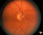
Vascular Disc Anomalies - Prepapillary Arterial Loop | Prepapillary arterial loop arising typically from the inferior retinal arterioles and projecting forward into the vitreous. 42 year old patient. Anatomy: Optic disc. Pathology: Congenital prepapillary arterial loop. Disease/Diagnosis: Congenital prepapillary arterial loop. Clinical: Asymptomatic. | Image |
| 11 |
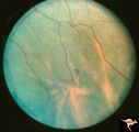 |
Von Hippel Lindau Disease | Von Hippel Lindau Disease with a mini retinal tumor. Pair with R1_C4b. Anatomy: Retina. Pathology: Hemangioblastoma. Disease/Diagnosis: Von Hippel Lindau disease. Clinical: No visual symptoms. Imaging: Flourescien angiogram in R1_C4b. | Image |
| 12 |
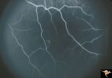 |
Von Hippel Lindau Disease | Von Hippel Lindau Disease with a mini retinal tumor. Flourescien angiogram shows how small tumor is. Pair with R1_C4a. Anatomy: Retina. Pathology: Hemangioblastoma. Disease/Diagnosis: Von Hippel Lindau disease. Clinical: No visual symptoms. Imaging: Flourescien angiogram. | Image |
| 13 |
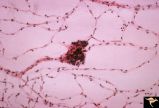 |
Von Hippel Lindau Disease Pathology of Mini Hemangioblastoma | Von Hippel Lindau Disease. Pathologic appearance of flat preparation of retina from necropsy study. Trypsin digestion study of retinal vascular bed with a mini VHL lesion. Anatomy: Retina. Pathology: Hemangioblastoma. Disease/Diagnosis: Von Hippel Lindau disease. Clinical: No visual symptoms. | Image |
| 14 |
 |
Von Hippel Lindau Disease with Photocoagulation Effect | Von Hippel Lindau Disease with appearance of Xenon photocoagulation on a mini hemangioblastoma. Anatomy: Retina. Pathology: Hemangioblastoma. Disease/Diagnosis: Von Hippel Lindau disease. Clinical: No visual symptoms. | Image |
1 - 25 of 14