Collection of materials relating to neuro-ophthalmology as part of the Neuro-Ophthalmology Virtual Education Library.
NOVEL: https://novel.utah.edu/
TO
- NOVEL724
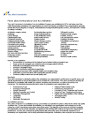
| Title | Creator | Description | Subject | ||
|---|---|---|---|---|---|
| 276 |
 |
Congenital and Secondary Syphilis | Gregory P. Van Stavern, MD | Images showing evideince of Congenital and Secondary Syphilis | Syphilis |
| 277 |
 |
Vision & Alzheimer's Disease | Victoria S. Pelak, MD | Alzheimer's Disease (AD) is an age-related neurodegenerative disorder with progressive loss of cognitive function over time. A clinical diagnosis for Probable AD Dementia requires the following: a loss of cognitive function in two or more cognitive domains (or in one cognitive domain along with a ch... | Vision; Alzheimer's Disease |
| 278 |
 |
White Dot Syndromes: MEWDS, AZOOR, AIBSE | Gregory P. Van Stavern, MD | Some have lumped Multiple Evanescent White Dot Syndrome (MEWDS), Acute Idiopathic Blind Spot Enlargement (AIBSE) with acute macular neuroretinopathy, and pseudo-presumed ocular histoplasmosis syndrome together with AZOOR (Acute Zonal Occult Outer Retinopathy). These conditions all present with visua... | White Dot Syndromes: MEWDS, AZOOR, AIBSE |
| 279 |
 |
Superonasal Transconjunctival Optic Nerve Sheath Decompression: A Modified Surgical Technique Without Extraocular Muscle Disinsertion | Kevin E. Lai, MD; Kenneth C. Lao, MD; Peter L. Hildebrand, MD; Bradley K. Farris, MD | Report on the surgical technique and outcomes of a modified medial transconjunctival approach to optic nerve sheath decompression (ONSD) in 15 patients. Supplemental Digital Content : Video that demonstrates the stONSD procedure. m4v: http://content.lib.utah.edu/cdm/ref/collection/EHSL-NOVEL/id/22... | Superonasal Transconjunctival Optic Nerve Sheath Decompression (ONSD); Surgical Technique |
| 280 |
 |
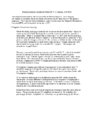
Disability Evaluation Under Social Security | John Pula, MD | A. How do we evaluate visual disorders? 1. What are visual disorders? Visual disorders are abnormalities of the eye, the optic nerve, the optic tracts, or the brain that may cause a loss of visual acuity or visual fields. A loss of visual acuity limits your ability to distinguish detail, read, or do... | Visual Impairment; Visual Disorders; Legal Blindness |
| 281 |
 |
Retinitis Pigmentosa - Rod Dystrophy | Gregory P. Van Stavern, MD | PowerPoint discussing retinitis pigmentosa, rod dystrophy. Retinitis Pigmentosa is a generalized retinal dystrophy with peripheral rather than central onset Primarily rod-cone dystrophy. Provides images. | Rod Dystrophy; Rod Dystrophy; Retinitis Pigmentosa; Night Dlindness |
| 282 |
 |
Tonic Pupil | Adesina, Ore-Ofe, MD | PowerPoint presentation covering tonic pupil, which is damage to ciliary ganglion or short posterior ciliary nerves. It causes denervation of the ciliary body and iris sphincter muscle. | Tonic Pupil |
| 283 |
 |
Horner's Carotid Dissection | Gregory P. Van Stavern, MD | PowerPoint describing Horner's Syndrome and Carotid Dissection. | Horner's Syndrome; Carotid Dissection; Dark Adaptation; Rod Dystrophy |
| 284 |
 |
Diffusion Weighted Imaging (DWI) | John Pula, MD | Diffusion weighted imaging sequences are often included as part of a routine brain MRI protocol. Imaging provides examples of DWI. | Diffusion Weighted Imaging; DWI |
| 285 |
 |
Photophobia for Patients - Large Print | Kathleen B. Digre, MD | The symptoms of light sensitivity are: an uncomfortable sense of brightness, squinting, frequent blinking, and redness of the eye (especially if the eye is dry). Involuntary eye closure and excessive blinking is seen with blepharospasm. Individuals will tend to seclude themselves in darkness. | Photophobia |
| 286 |
 |
Neuromyelitis Optica (NMO) | John Pula, MD | Slideshow describing condition. | Neuromyelitis Optica; NMO |
| 287 |
 |
Radiological Examination of the Visual System | John Pula, MD | An explanation of imaging types. | Visual System; Radiology; Imaging |
| 288 |
 |
Photophobia for Patients | Kathleen B. Digre, MD | The symptoms of light sensitivity are: an uncomfortable sense of brightness, squinting, frequent blinking, and redness of the eye (especially if the eye is dry). Involuntary eye closure and excessive blinking is seen with blepharospasm. Individuals will tend to seclude themselves in darkness. | Photophobia |
| 289 |
 |
Diffusion Tensor Imaging (DTI) | John Pula, MD | Diffusion tensor (DT) MRI applies the direction of water diffusion through tissues to map out neural pathways in the brain, such as white matter tracts. | Diffusion Tensor Imaging; DTI |
| 290 |
 |
Multiple Sclerosis Treatment Strategies | John Pula, MD | Slideshow exploring current treatment of multiple sclerosis. | Multiple Sclerosis; Multiple Sclerosis Treatment |
| 291 |
 |
Facts About Ambulatory Care Accreditation | Joint Commission on Accreditation of Healthcare Organizations (JCAHO) | The Joint Commission's Ambulatory Care Accreditation Program was established in 1975, and today more than 2,000 freestanding ambulatory care organizations are Joint Commission-accredited. These organizations generally fall into the broad categories of surgical, medical/dental and diagnostic/therapeu... | Ambulatory Care Accreditation |
| 292 |
 |
Branch Retinal Artery Occlusion with Multiple Retinal Emboli | Kathleen B. Digre, MD; James J. Corbett, MD | Slideshow describing condition. | Retinal Emboli; Emboli |
| 293 |
 |
Branch Retinal Vein Occlusion (BRVO) | Kathleen B. Digre, MD; James J. Corbett, MD | Slideshow describing condition. | Occlusion |
| 294 |
 |
Branch Retinal Artery Occlusion | Kathleen B. Digre, MD; James J. Corbett, MD | Slideshow describing condition. | Occlusion |
| 295 |
 |
Branch Retinal Emboli | Kathleen B. Digre, MD; James J. Corbett, MD | Slideshow describing condition. | Emboli |
| 296 |
 |
Calcific Emboli | Kathleen B. Digre, MD; James J. Corbett, MD | Slideshow describing condition. | Emboli |
| 297 |
 |
Central Retinal Vein Occlusion | Kathleen B. Digre, MD | Slideshow describing condition. | Occlusion |
| 298 |
 |
Craniopharyngioma and Optic Atrophy | Kathleen B. Digre, MD; James J. Corbett, MD | Slideshow describing condition. | Craniopharayngioma; Otpic Atrophy |
| 299 |
 |
CRAO with Ciliary Artery Sparing | Kathleen B. Digre, MD; James J. Corbett, MD | Slideshow describing condition. | CRAO |
| 300 |
 |
Central Cone Dystrophy Occult Macular Dystrophy | Gregory Van Stavern, MD | Slideshow describing condition of Central Cone Dystrophy Occult Macular Dystrophy | Central Cone Distrophy; Macular Dystrophy; Occult Macular Dystrophy |