A collection of videos relating to the diagnosis and treatment of eye movement disorders. This collection includes many demonstrations of examination techniques.
Dan Gold, D.O., Associate Professor of Neurology, Ophthalmology, Neurosurgery, Otolaryngology - Head & Neck Surgery, Emergency Medicine, and Medicine, The Johns Hopkins School of Medicine.
A collection of videos relating to the diagnosis and treatment of eye movement disorders.
NOVEL: https://novel.utah.edu/
TO
Filters: Collection: "ehsl_novel_gold"
| Title | Description | Type | ||
|---|---|---|---|---|
| 301 |
 |
Saccadic Pathways in the Brainstem and Cerebellum & Mechanism for Saccadic Dysmetria in Wallenberg Syndrome - Abnormal Function of the Brainstem/Cerebellar Saccadic Pathways with a Left Wallenberg Syndrome | The end result of a lesion involving the climbing fibers within the left lateral medulla is deficient rightward saccades (contralesional hypometric saccades), and over-active leftward saccades (ipsilesional hypermetric saccades), and ipsilesional ocular lateropulsion given this baseline imbalance. M... | Image |
| 302 |
 |
Saccadic Pathways in the Brainstem and Cerebellum & Mechanism for Saccadic Dysmetria in Wallenberg Syndrome - Normal Function of the Brainstem/Cerebellar Saccadic Pathways | The inferior cerebellar peduncle (ICP) carries climbing fibers to the dorsal vermis, and these fibers have an inhibitory influence over the Purkinje cells. These Purkinje cells normally inhibit the ipsilateral fastigial nucleus, and the fastigial nucleus projects to the contralateral inhibitory burs... | Image |
| 303 |
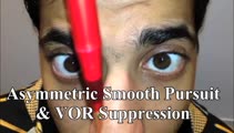 |
Saccadic Smooth Pursuit and Vestibulo-ocular Reflex Suppression (VORS) | 𝗢𝗿𝗶𝗴𝗶𝗻𝗮𝗹 𝗗𝗲𝘀𝗰𝗿𝗶𝗽𝘁𝗶𝗼𝗻: This is a 20-yo-man who suffered a left MCA stroke years prior. Upon evaluation of his eye movements, saccades and all classes of eye movements were normal, although his smooth pursuit and VORS were choppy to the left (ip... | Image/MovingImage |
| 304 |
 |
Sagging Eye Syndrome and Cerebellar Disease in Divergence Insufficiency | 𝗢𝗿𝗶𝗴𝗶𝗻𝗮𝗹 𝗗𝗲𝘀𝗰𝗿𝗶𝗽𝘁𝗶𝗼𝗻: This is a 70-year-old woman who presented with diplopia at distance. Her exam demonstrated orthophoria at near with a fairly comitant 8-10 PD esotropia at distance without abduction paresis, consistent with divergence ins... | Image/MovingImage |
| 305 |
 |
Sagittal Section of the Brainstem Showing Structures Related to Normal Eyelid Function | Seen here is a sagittal view of the brainstem, with the structures relevant to normal eyelid function highlighted. The M-group, which can be found medial to the riMLF (coordinates eye and lid movements), has (weak) projections to the facial nucleus for frontalis muscle contraction, and (strong) proj... | Image |
| 306 |
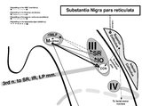 |
Sagittal Section of the Midbrain Showing Structures Related to Normal Eyelid Function | 𝗢𝗿𝗶𝗴𝗶𝗻𝗮𝗹 𝗗𝗲𝘀𝗰𝗿𝗶𝗽𝘁𝗶𝗼𝗻: During a vertical saccade, the rostral interstitial nucleus of the medial longitudinal fasciculus (riMLF) is activated, which excites the superior rectus (SR) and inferior oblique (IO) (IIIrd nerve) subnuclei. Additionall... | Image |
| 307 |
 |
Secondary Stroke Prevention | A brief overview of secondary stroke prevention. (TIA = Transient Ischemic Attack) | Text |
| 308 |
 |
Semicircular Pathways | Once the semicircular canal fibers leave the peripheral labyrinth, they synapse in the ipsilateral vestibular nucleus, and then ascend to the ocular motor nuclei. This enables the vestibulo-ocular reflex to respond to head movements in the plane of any canal or combination of canals. | Text |
| 309 |
 |
Sequelae of Cerebellar Hemorrhage - Gaze-evoked Nystagmus, Alternating Skew Deviation and Palatal Tremor | This is a 75-yo-woman presenting with a gait disorder. Two years prior, she suffered a cerebellar hemorrhage. On examination, there were typical cerebellar ocular motor signs including gaze-evoked nystagmus, choppy smooth pursuit and VOR suppression, and saccadic dysmetria. There was also an alterna... | Image/MovingImage |
| 310 |
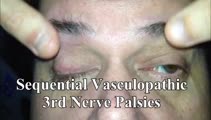 |
Sequential Vasculopathic 3rd Nerve Palsies with Preserved 4th Nerve Function | 65-yo-man with uncontrolled diabetes who developed sequential vasculopathic 3rd nerve palsies. In attempted downgaze, there's clear incyclotorsion OU suggestive of preserved 4th nerve function on both sides. There was complete recovery over months. Video shows bilateral 3rd nerve palsies with intact... | Image/MovingImage |
| 311 |
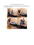 |
Short Canal Repositioning Maneuver for Anterior Canal BPPV | The Short Canal Repositioning Maneuver is used to treat anterior canal BPPV. 1. The patient's head is rotated 45-degrees towards the affected side. 2. The patient's maintains head in a 45-degree position and enters a head hanging position (40 degrees below the horizontal). 3. The patient then mainta... | Text |
| 312 |
 |
Short Canal Repositioning Maneuver for Anterior Canal BPPV (Video) | The Short Canal Repositioning Maneuver is used to treat anterior canal BPPV. 1. The patient's head is rotated 45-degrees towards the affected side. 2. The patient's maintains head in a 45-degree position and enters a head hanging position (40 degrees below the horizontal). 3. The patient then mainta... | Image/MovingImage |
| 313 |
 |
Side-lying Test for Right BPPV | The side-lying test is an alternative for the Dix Hallpike Test as it reduces the need for cervical extension. The interpretation of a positive test is the same as the Dix Hallpike Test. | Text |
| 314 |
 |
Side-lying Test for Right BPPV (Video) | The side-lying test is an alternative for the Dix Hallpike Test as it reduces the need for cervical extension. The interpretation of a positive test is the same as the Dix Hallpike Test. | Image/MovingImage |
| 315 |
 |
Sitting & Walking Oscillopsia in a Patient with Bilateral Vestibular Loss & Head Tremor | This is a 55-year-old man with oscillopsia for two reasons: He experienced oscillopsia at rest - so-called ‘sitting' oscillopsia - not from spontaneous nystagmus, but because of a combination of bilateral vestibular loss (BVL) and a mainly horizontal head tremor (this is sometimes referred to a... | Image/MovingImage |
| 316 |
 |
Skew Deviation and Spontaneous Nystagmus Due to Posterior Fossa Lesions | This is a 50-year-old woman who reported the abrupt onset of imbalance, right upper extremity incoordination and binocular vertical diplopia several months prior to her presentation to our clinic. On examination, she had a left hypertropia that was fairly comitant (measuring 5 prism diopters) assoc... | Image/MovingImage |
| 317 |
 |
Skew Deviation and the Triad of the Ocular Tilt Reaction (OTR) | This is a patient who presented with vertical diplopia, who was found to have a complete ocular tilt reaction including the following features: (1) Skew deviation - right hypertropia that was about 30 prism diopters in all directions of gaze including right, left, up, down, as well as in right and ... | Image/MovingImage |
| 318 |
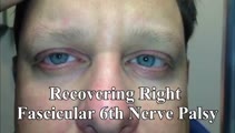 |
Slow Abducting Saccade in 6th Nerve Palsy | 40-yo-man with a right fascicular 6th nerve palsy due to stroke. There was improvement and only a minimal residual right abduction paresis OD by this visit, but still a relatively slow right abducting saccade seen in the video, especially apparent in the slow motion segment. Video shows slow abduct... | Image/MovingImage |
| 319 |
 |
Slow Horizontal, Vertical and Oblique Saccades in Spinocerebellar Ataxia Type I | This is a patient presenting with horizontal diplopia who was found to have divergence insufficiency, an esotropia greater at distance than near in the absence of abduction paresis. She also had very slow saccades, more so vertically than horizontally. This is particularly noticeable when asking h... | Image/MovingImage |
| 320 |
 |
Slow Horizontal, Vertical, Oblique Saccades and Gaze-evoked Nystagmus in Anti-AGNA-1 Encephalitis | This is a patient who presented subacutely with imbalance and dizziness. On examination, she had evidence of gaze evoked nystagmus, right internuclear ophthalmoplegia, as well as slow saccades horizontally and vertically. She was diagnosed with a rare antibody-mediated disorder, anti-AGNA-1 (antig... | Image/MovingImage |
| 321 |
 |
Slow Saccades Due to Unilateral Paramedian Pontine Reticular Formation (PPRF) Injury with Preserved Movements Using the Vestibulo-Ocular Reflex | 𝗢𝗿𝗶𝗴𝗶𝗻𝗮𝗹 𝗗𝗲𝘀𝗰𝗿𝗶𝗽𝘁𝗶𝗼𝗻: This is a 60-year-old man who presented for imbalance and oscillopsia 10 months after surgery and 8 months after radiation for Merkel cell carcinoma of the neck. He developed imbalance after surgery and diplopia and osci... | Image/MovingImage |
| 322 |
 |
Slow Volitional Saccades and Poor Fast Phases to an Optokinetic Stimulus, with Preserved Head Impulse Testing | This is a 67-year-old woman presenting with imbalance and binocular horizontal diplopia at near. On examination there were frequent square wave jerks, limited supraduction OU and convergence insufficiency, which explained her diplopia. Pursuit and suppression of the vestibulo-ocular reflex were sa... | Image/MovingImage |
| 323 |
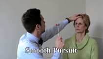 |
Smooth Pursuit | 𝗢𝗿𝗶𝗴𝗶𝗻𝗮𝗹 𝗗𝗲𝘀𝗰𝗿𝗶𝗽𝘁𝗶𝗼𝗻: A pursuit deficit in one direction suggests an ipsilesional localization, but beware of a superimposed spontaneous nystagmus; a pursuit deficit in all directions is commonly seen with cerebellar lesions. 𝗡𝗲𝘂𝗿�... | Image/MovingImage |
| 324 |
 |
Smooth Pursuit | Smooth pursuit: instruct the patient to hold their head steady, fix their eyes on the camera and slowly move the camera in the horizontal and vertical planes. Or, have the patient focus on their outstretched thumbnail (or other small fixation target), while following the slowly moving object horizon... | Image/MovingImage |
| 325 |
 |
Spinocerebellar Ataxia Type 3 with Gaze-Evoked Nystagmus and Bilateral Vestibular Loss | 𝗢𝗿𝗶𝗴𝗶𝗻𝗮𝗹 𝗗𝗲𝘀𝗰𝗿𝗶𝗽𝘁𝗶𝗼𝗻: This is a 50-year-old woman with an established diagnosis of spinocerebellar ataxia type 3 (SCA 3) with severe imbalance and head movement-induced oscillopsia. On examination, she had 1) bilateral vestibular loss (BVL) de... | Image/MovingImage |
