A collection of videos relating to the diagnosis and treatment of eye movement disorders. This collection includes many demonstrations of examination techniques.
Dan Gold, D.O., Associate Professor of Neurology, Ophthalmology, Neurosurgery, Otolaryngology - Head & Neck Surgery, Emergency Medicine, and Medicine, The Johns Hopkins School of Medicine.
A collection of videos relating to the diagnosis and treatment of eye movement disorders.
NOVEL: https://novel.utah.edu/
TO
Filters: Collection: "ehsl_novel_gold"
| Title | Description | Type | ||
|---|---|---|---|---|
| 351 |
 |
Test Your Knowledge - The Acute Vestibular Syndrome and Ptosis | What is the most likely localization in this patient presenting with vertical diplopia and acute onset prolonged vertigo? A. Right medial longitudinal fasciculus (MLF) B. Left medial longitudinal fasciculus C. Right medulla D. Left medulla E. Left midbrain A. Incorrect. A right MLF lesion (stroke, M... | Image/MovingImage |
| 352 |
|
Test Your Knowledge - Vertical Saccadic Palsy Due to Bilateral riMLF Infarctions | This is a 30-year-old who was found minimally responsive on the lounge floor of an ice skating rink. He was brought to the ED, where he had a GCS score of 8 (where 15 is normal) for poor responsiveness. His ocular motor exam is shown in the video. Regarding Finding #1, which of the following is fals... | Image/MovingImage |
| 353 |
|
Test Your Knowledge - Vertical-Torsional Nystagmus | Question #1: Watch the first portion of the video until you are told to stop. Is this vestibular nystagmus more likely to be peripheral or central? A. Peripheral B. Central Answer for #1: A. Incorrect. While the patient has upbeat-torsional (top poles beating toward the right ear) nystagmus which is... | Image/MovingImage |
| 354 |
|
Test Your Knowledge: The Acute Vestibular Syndrome with Gaze-Evoked Nystagmus and Bilaterally Abnormal Head Impulse Testing Due to Middle Cerebellar Peduncle and Flocculus Hemorrhage | This is a 70-year-old woman with a history of atrial fibrillation on warfarin presenting with acute prolonged vertigo and imbalance. In addition to the findings demonstrated in the first part of the video, what else should be seen to reassure the examiner that the etiology of her vertigo is benign? ... | Image/MovingImage |
| 355 |
 |
Testing for Adduction Lag in Partial INO Using an Optokinetic Stimulus | In this patient we demonstrate the use of an optokinetic stimulus to elicit an internuclear ophthalmoplegia (INO). Occasionally adduction appears to be normal with an INO, and an adduction lag with horizontal saccades should be sought as a confirmatory sign. Optokinetic tape is an easy way to assess... | Image/MovingImage |
| 356 |
|
Third and Sixth Nerve Palsies Due to Cavernous Sinus Meningioma | This is a 60-year-old woman with a large meningioma that was compressing the right cavernous sinus. Examination demonstrated a pupil-involving right 3rd nerve palsy with near complete external ophthalmoplegia (involving levator palpebrae, medial rectus, superior rectus, inferior rectus). There was a... | Image/MovingImage |
| 357 |
|
Third Nerve Palsy Due to Tolosa Hunt Syndrome | This is a 20-year-old woman presenting with severe left eye pain and diplopia for several days. Examination was consistent with a pupil sparing left 3rd nerve palsy with complete external ophthalmoplegia (involving levator palpebrae, medial rectus, superior rectus, inferior rectus). Abduction OS was... | Image/MovingImage |
| 358 |
 |
Torsional Jerk Nystagmus | Presented here are 3 patients with torsional jerk nystagmus. The first patient presented with vertigo and experienced oscillopsia due to her torsional nystagmus. Pure or predominantly torsional nystagmus is highly suggestive of a central process. Her nystagmus was unidirectional and followed Alexand... | Image/MovingImage |
| 359 |
 |
Torsional Nystagmus Due to Medullary Pilocytic Astrocytoma | 𝗢𝗿𝗶𝗴𝗶𝗻𝗮𝗹 𝗗𝗲𝘀𝗰𝗿𝗶𝗽𝘁𝗶𝗼𝗻: This is a 30-year-old woman who experienced headaches which led to an MRI and the diagnosis of a right medullary pilocytic astrocytoma, confirmed pathologically. Examination was performed a year after the initial diagnosi... | Image/MovingImage |
| 360 |
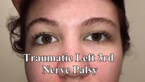 |
Traumatic 3rd Nerve Palsy with Aberrant Regeneration | 𝗢𝗿𝗶𝗴𝗶𝗻𝗮𝗹 𝗗𝗲𝘀𝗰𝗿𝗶𝗽𝘁𝗶𝗼𝗻: This is a 20-yo-woman who experienced severe head trauma and diplopia upon awakening from a coma several weeks after the injury. She had a partial left 3rd nerve palsy (adduction spared), and when she looked to the right ... | Image/MovingImage |
| 361 |
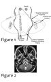 |
Triangle of Guillain-Mollaret | Seen here is a schematic representation of the Gullain-Mollaret triangle (Figure 1), also referred to as the dentato-olivary pathway, reflecting the 3 points of this imaginary triangle - 1) dentate nucleus, 2) red nucleus, and 3) inferior olivary nucleus. The olive sends decussating climbing fibers ... | Image |
| 362 |
 |
Trigeminal Motor Neuropathy with Weakness and Atrophy of the Muscles of Mastication | This is a man who was diagnosed with polio in childhood, which involved the motor (VIII) division of the right trigeminal nerve. The motor portion of the trigeminal nerve innervates the muscles of mastication (temporalis, masseter - both of which demonstrate wasting in this patient - as well as the ... | Image/MovingImage |
| 363 |
|
Trigeminal Neuropathy with Loss of the Corneal Reflex | This is a woman who underwent radiofrequency ablation for left trigeminal neuralgia. Examination demonstrated loss of facial sensation on the left in addition to an absent corneal reflex on the left, consistent with involvement of the V1 (ophthalmic) branch of the trigeminal nerve. When the cornea i... | Image/MovingImage |
| 364 |
 |
Trigeminal, Facial (with Aberrant Regeneration), and Vestibulocochlear Nerve Palsies Following Tumor Resection | This is a 30-yo-woman who underwent resection of a right trigeminal schwannoma. Post-operatively, she was vertiginous with a clearly + head impulse test to the right (and spontaneous left-beating nystagmus), had lost hearing in the right ear, had no facial sensation on the right, and had a right low... | Image/MovingImage |
| 365 |
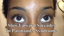 |
Two Patients with Parinaud's Syndrome with Slow Upward Saccades and Normal Upward Range of Movements | Presented here are two patients with Parinaud's syndrome: Patient 1) suffered a hemorrhage of the dorsal midbrain causing slow upward saccades (with convergence retraction nystagmus, but normal vertical range of eye movements), and light-near dissociation, and Patient 2) had a germinoma of the dorsa... | Image/MovingImage |
| 366 |
 |
Typical Features of Duane Syndrome Type 1 | 𝗢𝗿𝗶𝗴𝗶𝗻𝗮𝗹 𝗗𝗲𝘀𝗰𝗿𝗶𝗽𝘁𝗶𝗼𝗻: This is a patient seen for vestibular complaints, who on exam, was found to have (unrelated to her vestibular symptoms) impaired abduction OS. In adduction, there was narrowing of the palpebral fissure OS, a result of glo... | Image/MovingImage |
| 367 |
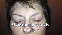 |
Typical Lid Signs (Cogan's Lid Twitch, Lid Hopping, Enhanced Ptosis) in Myasthenia Gravis | 𝗢𝗿𝗶𝗴𝗶𝗻𝗮𝗹 𝗗𝗲𝘀𝗰𝗿𝗶𝗽𝘁𝗶𝗼𝗻: This is a 60-yo-woman with MG who displays typical eyelid signs including Cogan's lid twitch, lid hopping (appreciated during horizontal smooth pursuit in this patient), and enhanced ptosis in accordance with Hering's law... | Image/MovingImage |
| 368 |
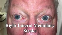 |
Unidirectional Nystagmus in Lateral Medullary Syndrome | This is a 70-yo-man who presented with acute vertigo. Examination demonstrated very mild spontaneous torsional nystagmus (towards the right ear) in primary (not seen well in this video), with robust downbeat-torsional (towards right ear) nystagmus in right gaze and (less robust) almost pure torsiona... | Image/MovingImage |
| 369 |
|
Unidirectional Nystagmus Two Days After Onset of Vestibular Neuritis | Image/MovingImage | |
| 370 |
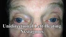 |
Unidirectional Vestibular Nystagmus | 60-yo-man with recurrent vertigo attacks - this video was taken during one of his typical attacks, and shows left-beating nystagmus that stayed left-beating in all directions of gaze, more in left gaze (in accordance with Alexander's Law), and less in right gaze. This pattern is more commonly seen w... | Image/MovingImage |
| 371 |
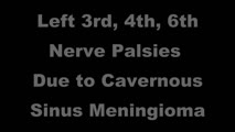 |
Unilateral 3rd, 4th, and 6th Nerve Palsies Due to Cavernous Sinus Meningioma | 𝗢𝗿𝗶𝗴𝗶𝗻𝗮𝗹 𝗗𝗲𝘀𝗰𝗿𝗶𝗽𝘁𝗶𝗼𝗻: This is a 50-year-old woman presenting with a partial 3rd nerve palsy (mild pupil involvement), partial 6th nerve palsy, and no clear incyclotorsion with downgaze, suggestive of additional 4th nerve palsy, all on the left... | Image/MovingImage |
| 372 |
 |
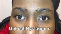
Upbeat and Downbeat Nystagmus Due to Anti-VGCC Antibodies | Seen here are two patients who presented with imbalance and vertical oscillopsia, the first with upbeat nystagmus, and the second with downbeat nystagmus. Both patients were found to have voltage-gated calcium channel antibodies in serum without evidence of systemic malignancy. The UBN patient had m... | Image/MovingImage |
| 373 |
|
Upbeat Nystagmus & Ocular Flutter Due to Cerebellar Pilocytic Astrocytoma | This is a 20-year-old woman who was diagnosed with a cerebellar pilocytic astrocytoma at age 10 after presenting with severe headaches and hydrocephalus. She underwent incomplete resection and radiation therapy at that time. She experienced mild vertical oscillopsia in upgaze at baseline, and increa... | Image/MovingImage |
| 374 |
 |
Upbeat Transitioning to Downbeat Nystagmus in Wernicke's Encephalopathy | This is a 30-year-old man with a history of alcohol abuse who presented to the hospital with inability to walk after several weeks of heavy drinking and malnutrition. While in the hospital, he noted that when he would look straight ahead, everything he saw would appear to be bouncing up and down - a... | Image/MovingImage |
| 375 |
 |
Upbeating and Gaze-evoked Nystagmus, V-pattern Esotropia from Bilateral 4th Nerve Palsies | Video example of a patient with upbeating and gaze-evoked nystagmus, V-pattern esotropia from bilateral 4th nerve palsies. | Image/MovingImage |