Collection of materials relating to neuro-ophthalmology as part of the Neuro-Ophthalmology Virtual Education Library.
NOVEL: https://novel.utah.edu/
TO
- NOVEL725
| Title | Creator | Description | Subject | ||
|---|---|---|---|---|---|
| 51 |
 |
A Case Series of Mydriasis from an Anticholinergic Antiperspirant | Aileen Antonio, MD; Inna Bondira, DO; Cameron Holicki, DO; Christopher Glisson, DO; Tatiana Deveney, MD; Lina Nagia, DO | Causes of anisocoria span a wide range, from benign to life-threatening, making it a common indication for Neuro-Ophthalmology referrals. One such cause is related to pharmacologic mydriasis due to direct or systemic exposure. We present a case series of four patients with different presentations of... | Anisocoria; Mydriasis; Pharmacologic Anisocoria; Anticholinergic Antiperspirant |
| 52 |
 |
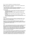
Anatomic and Physiologic Basis for Gaze Stability | Ariel Winnick and Meagan Seay, DO | Diagram describing the anatomic and physiologic basis of gaze stability. | Gaze Stability |
| 53 |
 |
Vitamin B12 Deficiency and Neuro-Ophthalmic Manifestations | Jourdan Carroll; Devin D. Mackay, MD | This presentation covers vitamin B12 deficiency, including etiology, signs and symptoms, neurologic and ophthalmic findings, a case presentation and treatment. | Vitamin B12 Deficiency and Neuro-Ophthalmic Manifestations |
| 54 |
 |
Optical Coherence Tomography (OCT) | Gunnar J. Goebel; Devin D. Mackay, MD | Introduction to OCT, including history, principles, interpretation, and applications. | Optical Coherence Tomography (OCT) |
| 55 |
 |
How to Find Funding Sources | Elizabeth Frakes, MLIS, AHIP | Brief description of funding sources and how to find them. | Funding |
| 56 |
 |
How to Submit an IRB Application | Elizabeth Frakes, MLIS, AHIP | A general outline of the steps involved in submitting an IRB application for research. | Institutional Review Board (IRB) |
| 57 |
 |
A Macro Dilemma: Demonstrations of the Anterior Chiasmal Syndrome and Wilbrand's Knee | Ariel Axelbaum, MD; Nurhan Torun, MD | Presentation reviewing two cases that demonstrate several cases of anterior chiasmal syndromes and the variability in patient presentations with sellar masses. | Anterior Chiasmal Syndromes; Masses of the Sella |
| 58 |
 |
Gerstmann Syndrome | Nicholas Bontrager; Padmaja Sudhakar, MD | Gerstmann syndrome refers to a constellation of four neurologic deficits: agraphia, acalculia, finger agnosia , and left-right disorientation. All symptoms must be present for a diagnosis of true Gerstmann syndrome. | Gerstmann Syndrome |
| 59 |
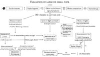 |
Pupil Evaluation Flowchart | Ariel Winnick; Eric Caskey, MD; Meagan Seay, DO | Flow chart outlining the evaluation of large or small pupils. | Pupil Evaluation |
| 60 |
 |
Neuro-Ophthalmic Manifestations of Mitochondrial Disease | Abhimanyu S. Ahuja, BS; Sidney M. Gospe III, MD, PhD | Classically, mitochondrial diseases exhibit a maternal inheritance pattern because pathogenic mutations in mitochondrial DNA (mtDNA) are transmitted exclusively via the maternal lineage due to rapid degradation of sperm-derived mitochondria early in embryogenesis. However, mutations in mtDNA may occ... | Mitochondrial Disease; Pathophysiology |
| 61 |
 |
Non-Surgical Management of Strabismus | Alex Christoff, MD | An overview of non-surgical treatment of strabismus. | Strabismus |
| 62 |
 |
Decompensated Phoria | Alex Christoff, MD | An overview of decompensated phoria and its treatment. | Decompensated Phoria |
| 63 |
 |
Susac's Syndrome | David O. Sohutskay; Kevin E. Lai, MD; Linda S. Williams, MD; Devin D. Mackay, MD | Overview of a case of Susac Syndrome. | Susac Syndrome |
| 64 |
 |
Anatomy of the Oculomotor Nerve (CN III) | Lucas E. Morgan, MS4; Nicholas A. Koontz, MD; Devin D. Mackay, MD | A detailed overview of the anatomic course of CN III, including a detailed pathway description and labeled MRI images, gross anatomy pictures, and structural models. | CN III; Third Cranial Nerve; Oculomotor Nerve; Anatomy; MRI |
| 65 |
 |
Cotton Wool Spots: The Basics | Arnav Gupta, BHSc; Rahul Sharma, MD, MPH | A presentation describing cotton wool spots, an abnormal finding on funduscopic exam of the retina of the eye. | Cotton Wool Spots; Retina |
| 66 |
 |
Disc Cupping: The Basics | Arnav Gupta, BHSc; Rahul Sharma, MD, MPH | A presentation describing optic disc cupping, due to damage of optic nerve fibres. | Disc Cupping; Optic Disc |
| 67 |
 |
Retinal Detachment: The Basics | Arnav Gupta, BHSc; Rahul Sharma, MD, MPH | A presentation describing retinal detachment. | Retinal Detachment |
| 68 |
 |
Retinal Exudate: The Basics | Arnav Gupta, BHSc; Rahul Sharma, MD, MPH | A presentation describing retinal exudate. | Retinal Exudate |
| 69 |
 |
Retinal Hemorrhage: The Basics | Arnav Gupta, BHSc; Rahul Sharma, MD, MPH | A presentation describing retinal hemorrhage. | Retinal Hemorrhage |
| 70 |
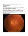 |
Situs Inversus Optic Disc Anomaly | Michael Hii, Medical Student; Ryan Walsh, MD | This patient was incidentally-noted to have anomalous appearance of the optic discs, right more so than left, consistent with situs inversus optic disc anomaly. She did not have any visual deficits related to this exam finding. ; The patient's fundus photos demonstrate situs inversus of the optic ... | Situs Inversus Optic Disc Anomaly |
| 71 |
 |
Age-related Macular Degeneration: The Basics | Arnav Gupta, BHSc; Rahul Sharma, MD, MPH | A presentation covering age-related macular degeneration ("ARMD" or "AMD"), an acquired, progressive, chronic, degenerative disease of the retina. | Macular Degeneration |
| 72 |
 |
Progressive Supranuclear Palsy (PSP) | Molly Cincotta, MD; Ali G. Hamedani, MD, MHS | Objectives:; To provide an overview of PSP and its pathophysiology;; To present typical clinical features of the disease with a focus on ocular findings;; To provide a template for work up, diagnosis and treatment; ; To demonstrate typical eye movement abnormalities seen in PSP | Progressive Supranuclear Palsy (PSP) |
| 73 |
 |
Computed Tomography (CT): Principles, Technique, and Neuro-ophthalmic Applications | Alex Fraser, MD | Presentation covering Computed Tomography principles, adverse effects, comparison vs. MRI, and assorted examples of neuro-ophthalmic interest. | Computed Tomography (CT) |
| 74 |
 |
Examining the Pediatric Patient for Non-Neuro-ophthalmologists | John Pula, MD | Ten need-to-know pearls for examining the pediatric patient, for non-neuro-ophthalmologists. | Pediatric Patient Exam |
| 75 |
 |
Examining the Comatose Patient for Non-Neuro-ophthalmologists | John Pula, MD | Seven need-to-know pearls for examining the pediatric patient, for non-neuro-ophthalmologists. | Comatose Patient Exam |