Best known for his world-renowned neuro-ophthalmology unit based at the University of California, San Francisco, William Hoyt, MD collected here more than 850 of his best images covering a wide range of disorders.
William F. Hoyt, MD, Professor Emeritus of Ophthalmology, Neurology and Neurosurgery, Department of Ophthalmology, University of California, San Francisco.
NOVEL: https://novel.utah.edu/
TO
Filters: Date: "1978" Collection: "ehsl_novel_wfh"
1 - 25 of 15
| Title | Description | Type | ||
|---|---|---|---|---|
| 1 |
 |
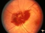
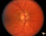
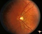
A303 Disc Swelling, Chorioretinal Disease | Neovascular net. Disc swelling with peripapillary neo-vascularization with subretinal hemorrhage. Anatomy: Optic disc; Retina. | Image |
| 2 |
 |
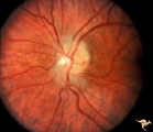
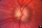
A404 Disc Swelling, Vitreous Effects | Disc elevation (swelling) and vitritis. Posterior vitreous detachment with vitritis. Incidental choroidal nevus. Anatomy: Optic disc; Vitreous; Retina. Pathology: Posterior vitreal detachment, disc swelling, and vitritis-horoidal nevus. Disease/ Diagnosis: Vitreal detachment from optic disc; choroid... | Image |
| 3 |
 |
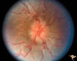
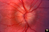
A407 Disc Swelling, Vitreous Effects | Prepapillary hemorrhage. Partial posterior vitreous detachment in myopic patient. Reference: Katz B, Hoyt WF. Intrapapillary and peripapillary hemorrhage in young patients with incomplete posterior vitreous detachment. Signs of vitreopapillary traction. Ophthalmology. 1995 Feb;102(2):349-54. Anatomy... | Image |
| 4 |
 |
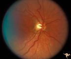
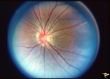
C16 Morning Glory Disc | "Morning Glory" disc. Note tapering edge pointing to basal encephalocele. Boy. Anatomy: Optic disc. | Image |
| 5 |
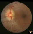 |
C303 Nodular Papillopathies (Sarcoid) | Lumpy infiltrative papillopathy in a patient with proven sarcoid. Anatomy: Optic disc. Pathology: Axoplasmic stasis due to sarcoid infiltration. Disease/ Diagnosis: Sarcoid papillopathy. Clinical: Unknown? | Image |
| 6 |
 |
E11 Disc Swelling with Central Vein Occlusion | Old retinal vein occlusion with optociliary bypass vessel at 3:00. Right eye. Anatomy: Optic disc; Retina. Pathology: Central retinal vein occlusion. Disease/ Diagnosis: Disc swelling due to central retinal vein occlusion. | Image |
| 7 |
 |
F202 Optic Nerve Sheath Meningioma | Optic nerve sheath meningioma. Note optociliary vein at 3:00. The disc is atrophic. Anatomy: Optic disc. Pathology: Chronic optic disc swelling caused by optic nerve sheath meningioma. Disease/ Diagnosis: Chronic optic disc swelling caused by optic nerve sheath meningioma. | Image |
| 8 |
 |
F2b06 Optic Disc Swelling from Optic Glioma | Right eye. Optic glioma with disc swelling. Anatomy: Optic disc. Pathology: Optic nerve glioma. Disease/ Diagnosis: Optic nerve swelling secondary to retrobulbar optic glioma. | Image |
| 9 |
 |
H86 Chiasmal Hemioptic Hypoplasia | Congenital bitemporal hemianopia with nasal hypoplasia. 24 year old man. Same patient as H_87. Anatomy: Optic disc. Pathology: Chiasmal hemioptic hypoplasia. Disease/ Diagnosis: Congenital anomaly involving chiasm. | Image |
| 10 |
 |
H87 Chiasmal Hemioptic Hypoplasia | Congenital bitemporal hemianopia with nasal hypoplasia. 24 year old man. Same patient as H_86. Anatomy: Optic disc. Pathology: Chiasmal hemioptic hypoplasia. Disease/ Diagnosis: Congenital anomaly involving chiasm. | Image |
| 11 |
 |
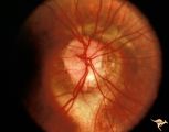
Hemorrhagic Complication of Drusen | PP31a, left and PP31, right taken in April. PP31c: left taken after an interval of 2 months. Hemorrhage. Hemorrhagic complications of drusen. 15 year old boy. Anatomy: Optic disc. Pathology: Drusen of the optic disc. Disease/Diagnosis: Drusen of the optic disc. Clinical: Patient complained of blurre... | Image |
| 12 |
 |
Hemorrhagic Complication of Drusen | PP31a, left and PP31, right taken in April. PP31c: left taken after an interval of 2 months. Hemorrhage. Hemorrhagic complications of drusen. 15 year old boy. Anatomy: Optic disc. Pathology: Drusen of the optic disc. Disease/Diagnosis: Drusen of the optic disc. Clinical: Hemorrhage in drusen disc. | Image |
| 13 |
 |
Hemorrhagic Complication of Drusen | PP31a, left and PP31, right taken in April. PP31c: left taken after an interval of 2 months. Hemorrhage. Hemorrhagic complications of drusen. 15 year old boy. Anatomy: Optic disc. Pathology: Drusen of the optic disc. Disease/Diagnosis: Drusen of the optic disc. Clinical: Drusen. | Image |
| 14 |
 |
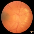
PP8a Crowded Disc with Significant Nasal Disc Blurring | Congenital nasal disc blurring. Myopic eyes. Thai girl patient. One wonders about vitreal adherence to the disc. PP 8a right eye. Pair with left eye in PP8b. Anatomy: Optic disc. Pathology: Normal variation of the optic disc. Disease/ Diagnosis: Normal variation of the optic disc. Congenital blurre... | Image |
| 15 |
 |
PP8b Crowded Disc with Significant Nasal Disc Blurring | Congenital nasal disc blurring. Myopic eyes. Thai girl patient. One wonders about vitreal adherence to the disc. PP 8b left eye. Pair with PP 8a right eye. Anatomy: Optic disc. Pathology: Normal variation of the optic disc. Disease/ Diagnosis: Normal variation of the optic disc. Congenital blurred d... | Image |
1 - 25 of 15