Collection of materials relating to neuro-ophthalmology as part of the Neuro-Ophthalmology Virtual Education Library.
NOVEL: https://novel.utah.edu/
TO
- NOVEL978
Filters: Collection: "ehsl_novel_novel"
| Title | Creator | Description | Subject | ||
|---|---|---|---|---|---|
| 201 |
 |
Grant Pieces - Research Grants | Silvia Sörensen, PhD | Lecture describing the parts of a research grant, including using human subjects. | Grant Writing |
| 202 |
 |
Protecting Human Subjects in Biomedical Research | Lisa R. Latchney, MS, CCRC | PowerPoint discussion of the history and development of ethics regulations in health research. | Ethical Issues in Research; Consent |
| 203 |
 |
Manuscripts: You Can Write These! | Elaine Smolock, PhD | Overview of writing techniques and parts of the manuscript, basic approach to writing results sections, what makes a good introduction, crafting a meaningful discussion, abstract and title suggestions, and how to get your editor's attention. | Writing Techniques |
| 204 |
 |
Acute Optic Neuritis | Neil R. Miller, MD, FACS | Overview of acute optic neuritis. | Optic Neuritis |
| 205 |
 |
Afferent Visual Pathway Disorders: Typical vs Atypical Optic Neuritis | Carmen Chan, FRCP, FRCOphth, FRCSEd(Ophth), FHKAM(Ophthalmology) | Discussion of typical vs atypical optic neuritis. | Optic Neuritis |
| 206 |
 |
Superior Segmental Optic Disc Hypoplasia (SSOH) "Topless Disc Syndrome" | Sparsh Jain, Medical Student; Ryan Walsh, MD | This is a case of superior segmental optic disc hypoplasia that was found incidentally after a screening visual field test revealed an asymptomatic inferior field defect in the left eye. The patient has a unilateral SSOH in the left eye. | Superior Segmental Optic Disc Hypoplasia (SSOH) |
| 207 |
 |
Bergmeister Papilla | Sumayya Almarzouqi, MD | A brief overview of Bergmeister papilla, a rare congenital disc anomaly. It arises from the center of the optic disc consists of a small tuft of fibrous tissue and represents a remnant of the fetal hyaloid artery. | Bergmeister Papilla |
| 208 |
 |
Modern Imaging of Optic Disc Drusen | Meagan Seay, DO | This is a short powerpoint describing imaging techniques (specifically OCT-EDI, fundus autofluorescence, and B-scan ultrasonography) for optic disc drusen. Examples of these techniques are included. | Optic Disc Drusen; Imaging; OCT-EDI; Fundus Autofluorescence; B-scan Ultrasonography |
| 209 |
 |
Suprasellar Meningioma | Sumayya Almarzouqi, MD | Description of a case of suprasellar or sellar mass causeing chiasmal compression. | Suprasellar Meningioma |
| 210 |
 |
Bony Anatomy of the Orbit | Aakash Patel; Amanda Henderson, MD | Narrated presentation on the bony anatomy of the orbit. | Bony Anatomy; Orbit |
| 211 |
 |
Ocular Surface, Cornea, & Lens | Sari Yordi, MD | Video lecture on the anatomy of the ocular surface, cornea, and lens. | Ocular Surface; Cornea; Lens |
| 212 |
 |
Lacrimal Pathways: Anatomy and Physiology | Sari Yordi, MD | Video lecture covering anatomy and physiology of the lacrimal pathways. | Lacrimal Pathways |
| 213 |
 |
Aqueous and Vitreous Humor | Sari Yordi, MD | Narrated lecture on the aqueous and vitreous humor. | Aqueous; Vitreous Humor |
| 214 |
 |
Physiology of Intraocular Pressure | Aakash Patel; Amanda Henderson, MD | Overview of the physiology of intraocular pressure. | Intraocular Pressure; Physiology |
| 215 |
 |
Arteriovenous Malformation | Justin Gibson, MD; Charles Prestigiacomo, MD | A diagnostic cerebroangiogram performed on a patient who presented with worst headache of life, found to have a Fisher Grade 3 subarachnoid hemorrhage. | Angiogram; Arteriovenous Malformation; AVM |
| 216 |
 |
Large Vessel Occlusion | Justin Gibson, MD; Charles Prestigiacomo, MD | Example of a diagnostic cerebroangiogram performed on a patient undergoing a stroke. | Angiogram; Stroke |
| 217 |
 |
Cerebral Aneurysm | Justin Gibson, MD; Charles Prestigiacomo, MD | Cerebral angiogram of a patient with an arteriovenous malformation, or AVM. | Angiogram; Cerebral Aneurysm |
| 218 |
 |
Normal Angiogram | Justin Gibson, MD; Charles Prestigiacomo, MD | Example of a normal diagnostic cerebroangiogram. | Angiogram |
| 219 |
 |
Anaesthesia for Eye Surgery and Associated Complications | Julie Smith, MBBS, FANZCA | Lecture covering commonly performed eye surgery and anaesthetic techniques. | Eye Surgery; Anesthesia |
| 220 |
 |
Neuroablative Procedures | Benjamin Jonker, MB BS, MMed(Clin Epi), FRACS | Video lecture covering neuro-ablative procedures that are relevant to neuro-ophthalmologists. | Ablative Procedures |
| 221 |
 |
Anaesthesia for Eye Surgery and Associated Complications Slides | Julie Smith, MBBS, FANZCA | Lecture covering commonly performed eye surgery and anaesthetic techniques. | Eye Surgery; Anesthesia |
| 222 |
 |
Embryology of the Eye | Yesha Shah, BSA, BBA; Amanda Henderson, MD | Video lecture covering the embryology of the eye. | Embryology; Eye |
| 223 |
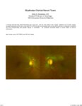 |
Myelinated Retinal Nerve Fibers | Scott N. Grossman, MD | A 33 year old man has noted chronically poor vision OS - left eye color noted to be 'orange' instead of red. fundus photos revealed myelinated retinal nerve fiber layer OU (OS>OD) with corresponding linear paracentral scotoma on Humphrey visual field 24-2 OS corresponding with greatest degree of my... | Myelinated Retinal Nerve Fibers |
| 224 |
 |
Cerebellar Anatomy on MRI | Joshua East, MD; Nicholas A. Koontz, MD; Devin D. Mackay, MD | Overview of structural anatomy of the cerebellum and surround structures on MRI images of the brain. | Cerebellar Anatomy; MRI |
| 225 |
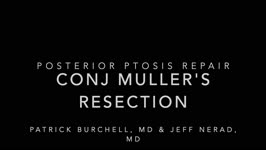 |
Conjunctiva Muller's Muscle Resection | Patrick Burchell, MD; Jeffrey Nerad, MD | Demonstration of conjunctiva Muller's muscle resection (CMMR). | Conjunctiva Muller's Muscle Resection; CMMR |