Best known for his world-renowned neuro-ophthalmology unit based at the University of California, San Francisco, William Hoyt, MD collected here more than 850 of his best images covering a wide range of disorders.
William F. Hoyt, MD, Professor Emeritus of Ophthalmology, Neurology and Neurosurgery, Department of Ophthalmology, University of California, San Francisco.
NOVEL: https://novel.utah.edu/
1 - 25 of 3
| Title | Description | Type | ||
|---|---|---|---|---|
| 1 |
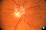 |
IA01 Atrophy with Optociliary Veins | 1994, perioptic nerve sheath meningioma, right eye, Optociliary vein dumping into disc edge at 4:00. Anatomy: Optic disc. Pathology: Optociliary vein. Disease/ Diagnosis: Perioptic nerve sheath meningioma. Clinical: Progressive visual loss | Image |
| 2 |
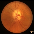 |
IA02 Atrophy with Optociliary Veins | 1971, left eye, perioptic nerve sheath meningioma, notice how vein dumps into adjacent choroid at 3:00. The darker venous blood can be seen at the disc edge. Anatomy: Optic disc. Pathology: Optociliary vein. Disease/ Diagnosis: Perioptic nerve sheath meningioma. Clinical: Progressive visual loss. | Image |
| 3 |
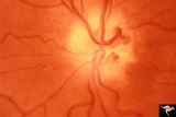 |
IA07 Atrophy with Optociliary Veins | Left eye, perioptic nerve sheath meningioma. Anatomy: Optic disc. Pathology: Optociliary vein. Disease/ Diagnosis: Perioptic nerve sheath meningioma. Clinical: Visual loss. | Image |
1 - 25 of 3