Collection of materials relating to neuro-ophthalmology as part of the Neuro-Ophthalmology Virtual Education Library.
NOVEL: https://novel.utah.edu/
TO
- NOVEL966
Filters: Collection: "ehsl_novel_novel"
| Title | Creator | Description | Subject | ||
|---|---|---|---|---|---|
| 1 |
 |
Branch Retinal Artery Occlusion with Multiple Retinal Emboli | Kathleen B. Digre, MD; James J. Corbett, MD | Slideshow describing condition. | Retinal Emboli; Emboli |
| 2 |
 |
Branch Retinal Vein Occlusion (BRVO) | Kathleen B. Digre, MD; James J. Corbett, MD | Slideshow describing condition. | Occlusion |
| 3 |
 |
Branch Retinal Artery Occlusion | Kathleen B. Digre, MD; James J. Corbett, MD | Slideshow describing condition. | Occlusion |
| 4 |
 |
Branch Retinal Emboli | Kathleen B. Digre, MD; James J. Corbett, MD | Slideshow describing condition. | Emboli |
| 5 |
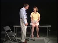
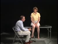
 |
Calcific Emboli | Kathleen B. Digre, MD; James J. Corbett, MD | Slideshow describing condition. | Emboli |
| 6 |
 |
Central/Branch Retinal Artery Occlusion (PowerPoint) | AAO/NANOS - American Academy of Ophthalmology / North American Neuro-Ophthalmology Society | Occlusion of a branch or central retinal artery may result in acute visual loss. The ophthalmoscopic findings are retinal whitening due to ischemic retina in the distribution of the occluded artery. Sparing or selective involvement of cilioretinal artery branches may occur. Patients with a central r... | Central/Branch Retinal Artery Occlusion |
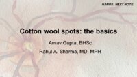
| 7 |
 |
Central Retinal Vein Occlusion | Kathleen B. Digre, MD | Slideshow describing condition. | Occlusion |
| 8 |
 |
Chest CT Thymoma (Guest Lecture) | Robert A. Novelline, MD | The patient is a 46 year old woman who presented in July 1977 with horizontal double vision lasting two weeks. Three weeks later the left upper eyelid started to droop and by the end of the day the eye was closed. She had no ptosis of the right eye and no generalized fatigue. She consulted an intern... | Unilateral Ptosis; Unilateral Lid Retraction; Myasthenic Lid Twitch; External Ophthalmoplegia; Ocular Myasthenia Gravis; Tensilon Test; Thymolipoma; Generalized Myasthenia Gravis; Unilateral Myasthenia Gravis; Myasthenic Ptosis; Lid Retraction; Lid Twitch |
| 9 |
 |
Chiasmal Herniation (PowerPoint) | AAO/NANOS - American Academy of Ophthalmology / North American Neuro-Ophthalmology Society | This woman was 61 years old when she underwent initial neuro-ophthalmologic evaluation. Twenty-three years earlier, she had undergone removal of a pituitary adenoma followed by radiation therapy. At that time, she had noted a preoperative visual field defect that improved somewhat after the surgery.... | Chiasmal Herniation |
| 10 |
 |
The Clinical Examination of Higher Order Visual Function: Syndrome-Based Approach | Victoria S. Pelak, MD; James R. Bateman, MD, MPH; Brianne Bettcher, PhD | Explanation of higher order visual function examination. See accompanying video, Double simultaneous visual field stimulation: https://collections.lib.utah.edu/ark:/87278/s6gn2h9d | Visual Function |
| 11 |
 |
The Clinical Examination of Higher Order Visual Function: Syndrome-based Approach - Visual Central Achromatopsia | Victoria S. Pelak, MD; James R. Bateman, MD, MPH; Brianne Bettcher, PhD | Explanation of higher order visual function examination. | Visual Function; Visual Central Achromatopsia |
| 12 |
 |
Clinical Characteristics of Ocular Lateropulsion | Jorge C Kattah, MD | A discussion of the normal mechanism that maintain the eyes in normal horizontal position. | Ocular Lateropulsion; Unilateral Gaze Palsy; Radiographic h-CGD |
| 13 |
 |
Clinical Visual Electrophysiology | Gregory P. Van Stavern, MD; Byron Lam, MD | A description of the use of electrophysiology to examine the visual system. | Electrophysiology; Visual Exam |
| 14 |
 |
Cogan's Lid Twitch Sign | Raed Behbehani, MD | Cogan's lid twitch sign is a twitch sign of he upper lid upon looking straight from a sustained downgaze position. It is associated with Ocular Myasthenia Gavis. | Myasthenia; Ptosis; Lid Twitch |
| 15 |
 |
Confrontation Visual Fields - A Concise Guide for Ophthalmology and Neurology Trainees | Stephen C. Pollock, MD | The guide describes the techniques required to competently perform confrontation visual fields. It outlines a basic screening protocol and discusses methods for further defining defects identified during the screening process. A mini-atlas of visual field defects is included as an appendix. | Confrontation Visual Fields; Visual Field Testing; Perimetry; Visual Field Loss; Visual Field Defect; Ocular Examination; Visual Sensory Evaluation; Neurologic Examination |
| 16 |
 |
Computed Tomography (CT) | Devin D. Mackay, MD | Explantation of computed tomography (CT) examinations. | Computed Tomography (CT) |
| 17 |
 |
Cone Dystrophy | Gregory P. Van Stavern, MD | PowerPoint discussing Cone Dystrophy: Early loss of central and color vision; Color impairment often out of proportion to loss of VA; Hemeralopia ("day blindness") prominent; Light sensitivity and photophobia; Macular changes variable, and may occur late- may "Bull's Eye" pattern; Abnormal Photost... | Cone Dystrophy; Occult Macular Dystrophy; Central Cone Dystrophy |
| 18 |
 |
Common Patterns of Visual Field Defects | Sean Gratton, MD; Sarah Lam, 6th year BA/MD | Lecture covering common visual field defects, including those of the retina, optic nerve, chiasm, and retrochiasmal. | Visual Field Defects |
| 19 |
 |
Complications of Strabismus Surgery and Botox | W. Walker Motley, MD | A narrated video slideshow outlining complications associated with strabismus surgery. | Strabismus; Surgery; Surgical Complications; Botox |
| 20 |
 |
Conjunctival Examination | Sovik De Sirkar, MSIII; Ore-ofe Adesina, MD, | Description of the conjunctival examination. | Conjunctival Examination |
| 21 |
 |
Contrast Sensitivity | Sean Gratton, MD | Explanation of contrast sensitivity. | Contrast Sensitivity |
| 22 |
 |
The Cornea and Scleral Examination | Sovik De Sirkar, MSIII; Ore-ofe Adesina, MD | Description of the cornea and scleral examination. | Cornea; Scleral Examination |
| 23 |
 |
Coordination Exam: Normal Exam: Finger-to-nose (includes Spanish audio & captions) | Paul D. Larsen, MD | The patient moves her pointer finger from her nose to the examiner's finger as the examiner moves his finger to new positions and tests accuracy at the furthest outreach of the arm. NeuroLogic Exam has been supported by a grant from the Slice of Life Development Fund at the University of Utah, the D... | Coordination Examination; Finger-to-nose Test |
| 24 |
 |
Coordination Exam: Normal Exam: Heel-to-shin (includes Spanish audio & captions) | Paul D. Larsen, MD | The patient places her heel on the opposite knee then runs the heel down the shin to the ankle and back to the knee in a smooth coordinated fashion. NeuroLogic Exam has been supported by a grant from the Slice of Life Development Fund at the University of Utah, the Department of Pediatrics and the O... | Coordination Examination; Heel-shin Test |
| 25 |
 |
Cotton Wool Spots: The Basics | Arnav Gupta, BHSc; Rahul Sharma, MD, MPH | A presentation describing cotton wool spots, an abnormal finding on funduscopic exam of the retina of the eye. | Cotton Wool Spots; Retina |