A collection of videos relating to the diagnosis and treatment of eye movement disorders. This collection includes many demonstrations of examination techniques.
Dan Gold, D.O., Associate Professor of Neurology, Ophthalmology, Neurosurgery, Otolaryngology - Head & Neck Surgery, Emergency Medicine, and Medicine, The Johns Hopkins School of Medicine.
A collection of videos relating to the diagnosis and treatment of eye movement disorders.
NOVEL: https://novel.utah.edu/
TO
Filters: Collection: "ehsl_novel_gold"
1 - 25 of 4
| Title | Description | Type | ||
|---|---|---|---|---|
| 1 |
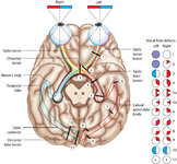 |
Figure 24: Typical Visual Field Defects Associated with Discrete Lesions Along the Visual Pathways | Specific monocular or binocular visual field defects can be highly localizing when the neuroanatomy of the visual pathways is understood. The temporal visual field corresponds to the nasal retina, while the nasal visual field corresponds to the temporal retina. 1) Left optic nerve lesion - while an ... | Image |
| 2 |
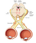 |
Figure 2: Parasympathetic Pathway for Pupillary Constriction | A bright light is shone in one eye, light enters the pupil and hyperpolarizes retinal photoreceptors which activates retinal ganglion cells. These signals propagate along the optic nerves, chiasm, optic tracts, and fibers responsible for the light reflex then synapse in the dorsal midbrain (prior to... | Image |
| 3 |
 |
Test Your Knowledge - Parinaud's Syndrome in Neurosarcoidosis | Watch the first segment of the video up to "Stop! What would you expect with vertical gaze?" and select the best response below. The patient also has mild right-beating nystagmus which can be ignored for the purposes of this question. A. The patient has pupillary findings consistent with bilateral 3... | Image/MovingImage |
| 4 |
 |
Expanded Nystagmus & Saccadic Intrusions/Oscillations Differential | Expanded nystagmus & saccadic intrusions/ oscillations differential | Text |
1 - 25 of 4