Collection of materials relating to neuro-ophthalmology as part of the Neuro-Ophthalmology Virtual Education Library.
NOVEL: https://novel.utah.edu/
TO
- NOVEL977
Filters: Collection: "ehsl_novel_novel"
| Title | Creator | Description | Subject | ||
|---|---|---|---|---|---|
| 526 |
 |
Usher Syndrome | Gregory P. Van Stavern, MD | Powerpoint describing Usher Syndrome, a hereditary condition characterized by congenital, bilateral, and profound sensorineural hearing loss, adolescent onset Retinitis Pigmentosa (RP) and vestibular areflexia | Usher Syndrome; Retinal Dystrophy; Retinitis Pigmentosa; Hearing Loss |
| 527 |
 |
Vision and Alzheimer's Disease | Victoria S. Pelak, MD | Slideshow describing condition. | Alzheimer's Disease |
| 528 |
 |
Visual Evoked Responses (Webvision) | Donnell J. Creel, MD, University of Utah | WebVision: The terms visually evoked potential (VEP), visually evoked response (VER) and visually evoked cortical potential (VECP) are equivalent. They refer to electrical potentials, initiated by brief visual stimuli, which are recorded from the scalp overlying visual cortex, VEP waveforms are ext... | Electrophysiology; Visual Evoked Responses |
| 529 |
 |
What is White? Normal White Structures | Gregory P. Van Stavern, MD | The only inherently "white" element in the normal eye is the sclera. | White in the Retina |
| 530 |
 |
2013 William F Hoyt Lecture: Neuro-Ophthalmology in Review: Around the Brain with 50 Fellows | Nancy J. Newman, MD | No matter what their ultimate specialty, every ophthalmologist needs to master the basics of neuroophthalmology. To that end, we must ensure that we continue to train effective teachers of neuro-ophthalmology. This is William F. Hoyt's most important lasting legacy and charge. In this same spirit, E... | History |
| 531 |
 |
Diffusion Weighted Imaging (DWI) | John Pula, MD | Diffusion weighted imaging sequences are often included as part of a routine brain MRI protocol. Imaging provides examples of DWI. | Diffusion Weighted Imaging; DWI |
| 532 |
 |
Diffusion Tensor Imaging (DTI) | John Pula, MD | Diffusion tensor (DT) MRI applies the direction of water diffusion through tissues to map out neural pathways in the brain, such as white matter tracts. | Diffusion Tensor Imaging; DTI |
| 533 |
 |
Multiple Sclerosis Treatment Strategies | John Pula, MD | Slideshow exploring current treatment of multiple sclerosis. | Multiple Sclerosis; Multiple Sclerosis Treatment |
| 534 |
 |
Facts About Ambulatory Care Accreditation | Joint Commission on Accreditation of Healthcare Organizations (JCAHO) | The Joint Commission's Ambulatory Care Accreditation Program was established in 1975, and today more than 2,000 freestanding ambulatory care organizations are Joint Commission-accredited. These organizations generally fall into the broad categories of surgical, medical/dental and diagnostic/therapeu... | Ambulatory Care Accreditation |
| 535 |
 |
Photophobia for Patients - Large Print | Kathleen B. Digre, MD | The symptoms of light sensitivity are: an uncomfortable sense of brightness, squinting, frequent blinking, and redness of the eye (especially if the eye is dry). Involuntary eye closure and excessive blinking is seen with blepharospasm. Individuals will tend to seclude themselves in darkness. | Photophobia |
| 536 |
 |
Neuromyelitis Optica (NMO) | John Pula, MD | Slideshow describing condition. | Neuromyelitis Optica; NMO |
| 537 |
 |
Radiological Examination of the Visual System | John Pula, MD | An explanation of imaging types. | Visual System; Radiology; Imaging |
| 538 |
 |
Photophobia for Patients | Kathleen B. Digre, MD | The symptoms of light sensitivity are: an uncomfortable sense of brightness, squinting, frequent blinking, and redness of the eye (especially if the eye is dry). Involuntary eye closure and excessive blinking is seen with blepharospasm. Individuals will tend to seclude themselves in darkness. | Photophobia |
| 539 |
 |
Branch Retinal Artery Occlusion with Multiple Retinal Emboli | Kathleen B. Digre, MD; James J. Corbett, MD | Slideshow describing condition. | Retinal Emboli; Emboli |
| 540 |
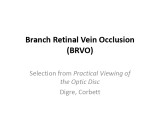 |
Branch Retinal Vein Occlusion (BRVO) | Kathleen B. Digre, MD; James J. Corbett, MD | Slideshow describing condition. | Occlusion |
| 541 |
 |
Branch Retinal Artery Occlusion | Kathleen B. Digre, MD; James J. Corbett, MD | Slideshow describing condition. | Occlusion |
| 542 |
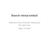 |
Branch Retinal Emboli | Kathleen B. Digre, MD; James J. Corbett, MD | Slideshow describing condition. | Emboli |
| 543 |
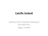 |
Calcific Emboli | Kathleen B. Digre, MD; James J. Corbett, MD | Slideshow describing condition. | Emboli |
| 544 |
 |
Central Retinal Vein Occlusion | Kathleen B. Digre, MD | Slideshow describing condition. | Occlusion |
| 545 |
 |
Craniopharyngioma and Optic Atrophy | Kathleen B. Digre, MD; James J. Corbett, MD | Slideshow describing condition. | Craniopharayngioma; Otpic Atrophy |
| 546 |
 |
CRAO with Ciliary Artery Sparing | Kathleen B. Digre, MD; James J. Corbett, MD | Slideshow describing condition. | CRAO |
| 547 |
 |
Central Cone Dystrophy Occult Macular Dystrophy | Gregory Van Stavern, MD | Slideshow describing condition of Central Cone Dystrophy Occult Macular Dystrophy | Central Cone Distrophy; Macular Dystrophy; Occult Macular Dystrophy |
| 548 |
 |
Pupillary reflex and the APD | Wade Crow, MD | Illustrations describing pupillary reflex. | Pupillary Reflex, APD |
| 549 |
 |
Vasospastic Amaurosis Fugax | Kathleen B. Digre, MD; James J. Corbett, MD | Slideshow describing condition. | Vasospastic Amaurosis Fugax |
| 550 |
 |
Hermann Ludwig Ferdinand von Helmholtz | Kathleen B. Digre, MD; James J. Corbett, MD | Biography of Hermann Ludwig Ferdinand von Helmholtz. | Hermann Ludwig Ferdinand von Helmholtz; History |