Best known for his world-renowned neuro-ophthalmology unit based at the University of California, San Francisco, William Hoyt, MD collected here more than 850 of his best images covering a wide range of disorders.
William F. Hoyt, MD, Professor Emeritus of Ophthalmology, Neurology and Neurosurgery, Department of Ophthalmology, University of California, San Francisco.
NOVEL: https://novel.utah.edu/
TO
Filters: Collection: "ehsl_novel_wfh"
| Title | Description | Type | ||
|---|---|---|---|---|
| 601 |
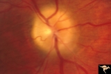 |
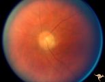
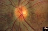
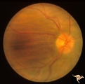
Papilledema due to Brain Tumor - Natural History | Right eye 3.5 years after presentation. Atrophy appears about the same. Note especially the narrowing of retinal arterioles. Visual loss is prfound in both eyes. Note the horizontal retinal folds. Papilledema due to brain tumor - 3 year natural history. Patient refused treatment. Man. | Image |
| 602 |
 |
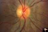
Papilledema due to Brain Tumor - Natural History | Left eye at 33 months after presentation. Atrophy appears about the same. Note especially the narrowing of retinal arterioles. Visual loss is severe in both eyes. Papilledema due to brain tumor - 3 year natural history. Patient refused treatment. Man. | Image |
| 603 |
 |
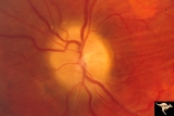
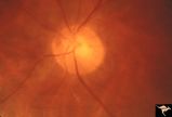
Papilledema with Choroidal Folds | Chronic papilledema with choroidal folds. Frontal astrocytoma. Man. Anatomy: Optic disc. Pathology: Papilledema. Disease/Diagnosis: Chronic papilledema. | Image |
| 604 |
 |
Papilledema with Choroidal Folds | Chronic papilledema with choroidal folds. Frontal astrocytoma. Flouroscein angiogram of choroidal folds. Man. | Image |
| 605 |
 |
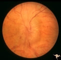
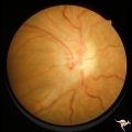
Paraneoplastic Retinopathy | Man with oat cell carcinoma of the lung with paraneoplastic retinopathy. Narrowed arterioles. Note absence of obvious retinal pigmentary degeneration. Cancer associated retinopathy syndrome (CAR Syndrome or Sawyer-Sellhorst Syndrome) (Ref: Sawyer, Sellhorst, Hoyt). Anatomy: Retina. Pathology: Oat c... | Image |
| 606 |
 |
Paraneoplastic Retinopathy | Man with oat cell carcinoma of the lung with paraneoplastic retinopathy. Narrowed arterioles. Note absence of obvious retinal pigmentary degeneration. Cancer associated retinopathy syndrome (CAR Syndrome or Sawyer-Sellhorst Syndrome) (Ref: Sawyer, Sellhorst, Hoyt). Anatomy: Retina. Pathology: Oat c... | Image |
| 607 |
 |
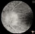
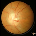
Paraneoplastic Retinopathy | Cancer associated retinopathy syndrome with extreme retino-arteriolar narrowing. CAR Syndrome. Anatomy: Retina. Pathology: Oat cell carcinoma of the lung with paraneoplastic retinopathy. Disease/Diagnosis: Cancer associated retinopathy. Clinical: Progressive visual loss, progressive night blindness,... | Image |
| 608 |
 |
Paraneoplastic Retinopathy | Cancer associated retinopathy syndrome with extreme retino-arteriolar narrowing. CAR Syndrome. Anatomy: Retina. Pathology: Oat cell carcinoma of the lung with paraneoplastic retinopathy. Disease/Diagnosis: Cancer associated retinopathy. Clinical: Progressive visual loss, progressive night blindness,... | Image |
| 609 |
 |
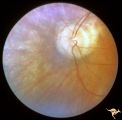
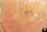
Pigment Epithelial Hamartoma of Optic Disc | Optic disc tumor discovered incidentally in a 32 year old Asian woman who had no complaints about visual function in her involved left eye. Fundus slide shows granular elevation of left disc obscurring major disc vessels. Some of the granules has a shiny crystalline appearance. Near the vessel entra... | Image |
| 610 |
 |
Pigmentary Retinopathy with Peripheral Neuropathy | Pigmentary retinopathy in a patient with peripheral neuropathy. Possible Refsums Syndrome. Optic disc shows some arteriole narrowing. Anatomy: Retina. Pathology: Peripheral nerve degeneration. Disease/Diagnosis: Retinitis pigmentosa with hereditary peripheral degeneration. Clinical: Blindness. | Image |
| 611 |
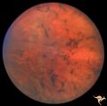 |
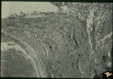
Pigmentary Retinopathy with Peripheral Neuropathy | Pigmentary retinopathy in a patient with peripheral neuropathy. Possible Refsums. Typical bone spicules in periphery. Anatomy: Retina. Pathology: Peripheral nerve degeneration. Disease/Diagnosis: Retinitis pigmentosa with hereditary peripheral degeneration. Clinical: Blindness. | Image |
| 612 |
 |
Pigmentary Retinopathy with Peripheral Neuropathy (Flecked Retinal Syndrome or Fundus Flavimaculatus) | Pigmentary retinopathy with peripheral neuropathy (Flecked Retinal Syndrome or Fundus Flavimaculatus) in a young woman. Anatomy: Retina. Pathology: Peripheral nerve degeneration. Disease/Diagnosis: Retinitis pigmentosa with hereditary peripheral degeneration. Clinical: Blindness. | Image |
| 613 |
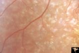 |
Pigmentary Retinopathy with Peripheral Neuropathy (Flecked Retinal Syndrome or Fundus Flavimaculatus) | Pigmentary retinopathy with peripheral neuropathy (Flecked Retinal Syndrome or Fundus Flavimaculatus) in a young woman. Anatomy: Retina. Pathology: Peripheral nerve degeneration. Disease/Diagnosis: Retinitis pigmentosa with hereditary peripheral degeneration. Clinical: Blindness. | Image |
| 614 |
 |
Pigmentary Retinopathy with Peripheral Neuropathy (Flecked Retinal Syndrome or Fundus Flavimaculatus) | Pigmentary retinopathy with peripheral neuropathy (Flecked Retinal Syndrome or Fundus Flavimaculatus) in a young woman. Anatomy: Retina. Pathology: Peripheral nerve degeneration. Disease/Diagnosis: Retinitis pigmentosa with hereditary peripheral degeneration. Clinical: Blindness. | Image |
| 615 |
 |
Pigmentary Retinopathy with Peripheral Neuropathy (Flecked Retinal Syndrome or Fundus Flavimaculatus) | Pigmentary retinopathy with peripheral neuropathy (Flecked Retinal Syndrome or Fundus Flavimaculatus) in a young woman. Anatomy: Retina. Pathology: Peripheral nerve degeneration. Disease/Diagnosis: Retinitis pigmentosa with hereditary peripheral degeneration. Clinical: Blindness. | Image |
| 616 |
 |
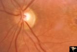
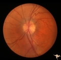
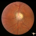
Post Papilledema | Right eye. Post Papilledema with minimal optic disc changes after treatment for temporal lobe glioma. Minimal optic disc haze. Optic disc. Pathology: Papilledema. Disease/Diagnosis: Post Papilledema due to temporal lobe glioma. | Image |
| 617 |
 |
Post Papilledema | Left eye. Post Papilledema with minimal optic disc changes after treatment for temporal lobe glioma. Minimal optic disc haze. | Image |
| 618 |
 |
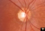
Post Papilledema Disc Blurring | Left eye. 8 year old boy. Post papilledema due to brain tumor. Note the entire peripapillary nerve fiber is blurred but the optic discs are barely elevated. Anatomy: Optic disc. Pathology: Brain tumor. Disease/Diagnosis: Papilledema. Clinical: Post papilledema due to brain tumor. | Image |
| 619 |
 |
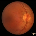
Post Papilledema Disc Blurring | Right eye. 8 year old boy. Post papilledema due to brain tumor. Note the entire peripapillary nerve fiber is blurred but the optic discs are barely elevated. Anatomy: Optic disc. Pathology: Post papilledema. Disease/Diagnosis: Post papilledema due to brain tumor. | Image |
| 620 |
 |
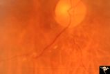
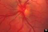
Post Papilledema Retinal Choroidal Bypass (Optociliary) | Right eye. Post papilledema retinal choroidal bypass (optociliary). Arterial venous malformation draining into saggital sinus causing papilledema and retinal choroidal collaterals. Anatomy: Optic disc. Pathology: Post papilledema. Disease/Diagnosis: Post papilledema with retinal choroidal bypass ves... | Image |
| 621 |
 |
Post Papilledema Retinal Choroidal Bypass (Optociliary) | Left eye. Post papilledema retinal choroidal bypass (optociliary). Arterial venous malformation draining into saggital sinus causing papilledema and retinal choroidal collaterals. Anatomy: Optic disc. Pathology: Post papilledema. Disease/Diagnosis: Post papilledema with retinal choroidal bypass vess... | Image |
| 622 |
 |
Post Papilledema with Choroidal Folds | Right eye. Post papilledema with choroidal folds due to brain tumor. Anatomy: Optic disc. Pathology: Post papilledema. Disease/Diagnosis: Post papilledema with choroidal folds. | Image |
| 623 |
 |
Post Papilledema, Secondary Optic Atrophy | Right eye. Post papilledema with chronic gliosis. arterial narrowing. ""high-water"" marks. Man. Anatomy: Optic disc. Pathology: Post papilledema. Disease/Diagnosis: Post papilledema with optic atrophy. | Image |
| 624 |
 |
Post Papilledema, Secondary Optic Atrophy | Left eye. Post papilledema with chronic gliosis. arterial narrowing. "high-water" marks. Man. Anatomy: Optic disc. Pathology: Post papilledema. Disease/Diagnosis: Post papilledema with optic atrophy. | Image |
| 625 |
 |
PP5b Crowded Disc | PP5a: left eye;PP5 b: left eye X 2 magnification; congenital disc blurring. Boy. Anatomy: Optic disc. Pathology: Normal variation of the optic disc. Disease/ Diagnosis: Normal variation of the optic disc. Congenital blurred disc. Clinical: Blurred disc margin. Beautiful example of difficult differen... | Image |