Best known for his world-renowned neuro-ophthalmology unit based at the University of California, San Francisco, William Hoyt, MD collected here more than 850 of his best images covering a wide range of disorders.
William F. Hoyt, MD, Professor Emeritus of Ophthalmology, Neurology and Neurosurgery, Department of Ophthalmology, University of California, San Francisco.
NOVEL: https://novel.utah.edu/
TO
Filters: Collection: "ehsl_novel_wfh"
| Title | Description | Type | ||
|---|---|---|---|---|
| 151 |
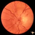 |
C205 Papillitis with Macular Star, Cat Scratch Disease | Proven Bartonella neuroretinitis. Macular star present, but not visible on image. 33 year old woman. Anatomy: Optic disc; Retina. Pathology: Axoplasmic stasis due to inflammation. Disease/ Diagnosis: Bartonella Henslae (Cat Scratch). Clinical: Visual blurring. | Image |
| 152 |
 |
C206 Papillitis with Macular Star, Cat Scratch Disease | Proven Bartonella neuroretinitis. Resolved papillitis with residual retinal exudate. Man. Anatomy: Optic disc; Retina. Pathology: Axoplasmic stasis due to inflammation. Disease/ Diagnosis: Bartonella Henslae (Cat Scratch). Clinical: Visual blurring; Ocular edema with macular star (ODEMS). | Image |
| 153 |
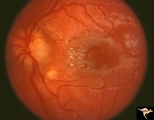 |
C207 Papillitis with Macular Star, Cat Scratch Disease | Proven Bartonella neuroretinitis. Anatomy: Optic disc; Retina. Pathology: Axoplasmic stasis due to inflammation. Disease/ Diagnosis: Bilateral Bartonella Henslae (Cat Scratch). Clinical: Visual blurring; Ocular disc edema with macular star (ODEMS). | Image |
| 154 |
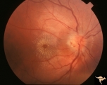 |
C208 Papillitis with Macular Star, Cat Scratch Disease | Proven Bartonella neuroretinitis. Man. Anatomy: Optic disc; Retinitis. Pathology: Axoplasmic stasis due to inflammation. Disease/ Diagnosis: Bartonella Henslae (Cat Scratch). Clinical: Visual blurring; Ocular disc edema with macular star (ODEMS). | Image |
| 155 |
 |
C209 Papillitis with Macular Star, Cat Scratch Disease | Proven Bartonella neuroretinitis. Woman. Anatomy: Optic disc; Retina. Pathology: Axoplasmic stasis due to inflammation. Disease/ Diagnosis: Bartonella Henslae (Cat Scratch). Clinical: Visual blurring without visual field defect; Ocular disc edema with macular star (ODEMS). | Image |
| 156 |
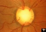 |
C21 Empty Disc | Right eye. All cilioretinal fundus. No central retinal artery. Handmann anomaly. Frequently associated with renal dysplasia. Pair with C_22 an C_23. Reference: Barroso LH, Hoyt WF, Narahara M. Can the arterial supply of the retina in man be exclusively cilioretinal? J Neuroophthalmol. 1994 Jun;14(2... | Image |
| 157 |
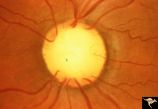 |
C22 Empty Disc | Left eye. All cilioretinal fundus. No central retinal artery. Handmann anomaly. Frequently associated with renal dysplasia. Pair with C_21 an C_23. Reference: Barroso LH, Hoyt WF, Narahara M. Can the arterial supply of the retina in man be exclusively cilioretinal? J Neuroophthalmol. 1994 Jun;14(2)... | Image |
| 158 |
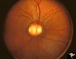 |
C23 Empty Disc | Father of patient in C_21 and C_22. Father has central retinal artery, multiple cilioretinal arteries and had previously unsuspected renal failure. Papillorenal Syndrome (PRS). Reference: Parsa,CF et al. Ophthalmology. 2001. 108(4): 738-49Barroso LH, Hoyt WF, Narahara M. Can the arterial supply of ... | Image |
| 159 |
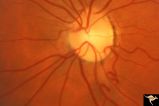 |
C24 Empty Disc | Left eye. Multiple cilioretinal arteries. Child of C_25 Anatomy: Optic disc. | Image |
| 160 |
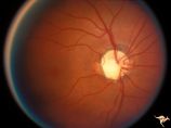 |
C25 Empty Disc | Right eye. Multiple cilioretinal arteries. Visual function normal. Father of C_24. Anatomy: Optic disc | Image |
| 161 |
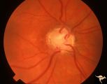 |
C26 Empty Disc | Right eye. Multiple cilioretinal arteries. Patient had dysplastic kidneys. Papillorenal Syndrome (PRS). Hand motion vision. 17 year old girl. Anatomy: Optic disc. | Image |
| 162 |
 |
C27 Empty Disc | Multiple cilioretinal arteries. Chronic interstitial nephritis. Renal and optic disc dysplasia. Papilorenal Syndrome (PRS). No central retinal artery. Anatomy: Optic disc. | Image |
| 163 |
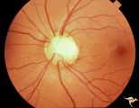 |
C28 Empty Disc | Left eye. Woman. Multiple cilioretinal vessels. Visual function normal. Anatomy: Optic disc. | Image |
| 164 |
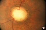 |
C29 Empty Disc | Left eye. Papilorenal Syndrome (PRS). Pair with C_30. Anatomy: Optic disc. | Image |
| 165 |
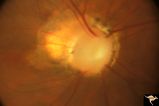 |
C30 Empty Disc | Right eye. Papillorenal Syndrome (PRS). Pair with C_29. Anatomy: Optic disc. | Image |
| 166 |
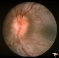 |
C301 Nodular Papillopathies (Sarcoid) | Disc swelling. Sarcoid papillopathy. Note infiltrative nodule at 9:00 on the disc.The patient had proven sarcoid. Perivenous inflammatory cuffing visible on image C3_02. Right eye. Pair with C3_02. Anatomy: Optic disc; Retina. Pathology: Axoplasmic stasis due to sarcoid infiltration. Disease/ Diagn... | Image |
| 167 |
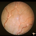 |
C302 Nodular Papillopathies (Sarcoid) | Perivenous Inflammatory Cuffing in a Patient with Proven Sarcoid. Left eye. Pair with C3_01. Anatomy: Retina. Pathology: Axoplasmic stasis due to sarcoid infiltration with retinal venous exudation? Disease/ Diagnosis: Sarcoid papillopathy with perivenous inflammatory disease. Clinical: Unknown? | Image |
| 168 |
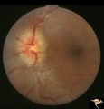 |
C303 Nodular Papillopathies (Sarcoid) | Lumpy infiltrative papillopathy in a patient with proven sarcoid. Anatomy: Optic disc. Pathology: Axoplasmic stasis due to sarcoid infiltration. Disease/ Diagnosis: Sarcoid papillopathy. Clinical: Unknown? | Image |
| 169 |
 |
C304 Nodular Papillopathies (Sarcoid) | Lumpy nodular disc infiltration from sarcoid. Anatomy: Optic disc. Pathology: Axoplasmic stasis due to sarcoid infiltration. Disease/ Diagnosis: Sarcoid papillopathy. Clinical: Unknown? | Image |
| 170 |
 |
C305 Nodular Papillopathies (Sarcoid) | July 1984 shows multiple infiltrative nodules on the optic disc in addition to circumferential subretinal yellow exudates. 32 year old black woman. Same patient as C3_06 and C3_07. Anatomy: Optic disc; Retina. Pathology: Axoplasmic stasis due to sarcoid infiltration and retinal exudation. Disease/ ... | Image |
| 171 |
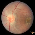 |
C306 Nodular Papillopathies (Sarcoid) | Lumpy disc swelling with retinal folds and a macular star in a patient with sarcoid. Presentation in October 1983. Same patient as C3_05 and C3_07. Anatomy: Optic disc; Retina. Pathology: Axoplasmic stasis due to sarcoid infiltration. Disease/ Diagnosis: Axoplasmic stasis due to sarcoid infiltration... | Image |
| 172 |
 |
C307 Nodular Papillopathies (Sarcoid) | Fluorescein angiogram shows striking staining of the sarcoid nodules. July 1984. Same patient as C3_05 and C3_06. Corresponds with July 1984 image, C3_06. Anatomy: Optic disc. Pathology: Axoplasmic stasis due to sarcoid infiltration. Disease/ Diagnosis: Sarcoid papillopathy. Clinical: Unknown? | Image |
| 173 |
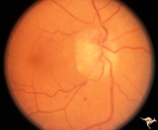 |
C308 Nodular Papillopathies (Sarcoid) | Nodular infiltrative papillopathy in a patient with sarcoid. Woman. Anatomy: Optic disc. Pathology: Axoplasmic stasis due to sarcoid infiltration. Disease/ Diagnosis: Sarcoid papillopahty. Clinical: Unknown? | Image |
| 174 |
 |
C31 Empty Disc | Right eye. Papillorenal Syndrome (PRS). Same patient as C_32. Anatomy: Optic disc. | Image |
| 175 |
 |
C32 Empty Disc | Left eye. Papillorenal Syndrome (PRS). Same patient as C_31. Anatomy: Optic disc. | Image |
