Collection of materials relating to neuro-ophthalmology as part of the Neuro-Ophthalmology Virtual Education Library.
NOVEL: https://novel.utah.edu/
TO
- NOVEL981
Filters: Collection: "ehsl_novel_novel"
| Title | Creator | Description | Subject | ||
|---|---|---|---|---|---|
| 101 |
 |
Sports Related Head Injuries | Jessica Darusz, MD; Sean Gratton, MD | This narrated PowerPoint reviews the basics of sports related head injuries. It emphasizes assessment tools and treatment decisions in assessing athletes with concussion and other head injuries. | Concussion; Sports-related Head Injuries; Post-concussion Syndrome |
| 102 |
 |
Postconcussion Syndrome and Postconcussion Headache | Jessica Darusz, MD; Sean Gratton, MD | This brief presentation describes the pathophysiology, evaluation, and management of concussion, with an emphasis on postconcussion headache. | Concussion; Postconcussion Syndrome; Postconcussion Headache |
| 103 |
 |
Morning Glory Disc Anomaly | Bashaer Aldhahwani, MD; Carlos Ernesto Mendoza Santiesteban, MD | A colored fundus photo of a patient with morning glory anomaly. Morning glory anomaly is a rare congenital malformation of the optic nerve. The morning glory disc anomaly can be seen with transsphenoidal basal encephalocele. It is known as morning glory syndrome when it is associated with systemic s... | Optic Disc Anomaly |
| 104 |
 |
Curtain Sign (Enhanced Ptosis) | Bashaer Aldhahwani, MD; Hong Jiang, MD, PhD | This is a 78-year-old male patient who presented with diplopia, right eyelid ptosis, and ophthalmoplegia. He had severe ptosis OD and pseudo-proptosis (lid retraction) OS at baseline, but when the right eyelid was manually elevated, there was marked enhanced ptosis of the left eyelid (Video). He was... | Myasthenia GravIs; Clinical Signs |
| 105 |
 |
Curtain Sign (Enhanced Ptosis) - Associated Image 1 | Bashaer Aldhahwani, MD; Hong Jiang, MD, PhD | This is a 78-year-old male patient who presented with diplopia, right eyelid ptosis, and ophthalmoplegia. He had severe ptosis OD and pseudo-proptosis (lid retraction) OS at baseline, but when the right eyelid was manually elevated, there was marked enhanced ptosis of the left eyelid (Video). He was... | Myasthenia GravIs; Clinical Signs |
| 106 |
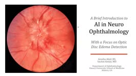 |
A Brief Introduction to AI in Neuro-ophthalmology | Areeba Abid, BS; Sachin Kedar, MD | In this video, we describe the basics of artificial intelligence and machine learning as applicable to clinical neuro-ophthalmologists. We use the example from a recent publication, where AI software was used to detect optic disc edema in fundus images. | Artificial Intelligence; Machine Learning |
| 107 |
 |
Optic Nerve Hypoplasia (ONH) - Double Ring Sign | Bashaer Aldhahwani, MD; Joshua Pasol, MD | Optic nerve hypoplasia (ONH) is characterized by a decreased number of optic nerve axons. It can present unilaterally or bilaterally, Isolated or associated with midline cerebral structural defects, such as septum pellucidum absence, agenesis of corpus callosum, cerebral hemisphere abnormalities, or... | Optic Nerve Hypoplasia (ONH) |
| 108 |
 |
Introduction to NANOS NOTE | Karl C. Golnik, MD | Introduction to NANOS NOTE, a resource for non-neuro-ophthalmologists describing common examination techniques. | Neuro-Ophthalmology Examination Techniques |
| 109 |
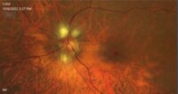 |
Myelinated Retinal Nerve Fiber Layer | Bashaer Aldhahwani, MD; Hong Jiang, MD, PhD | A 78 YOF with no visual symptoms has an incidental finding of yellow-white well-demarcated patches with ragged borders at the peripapillary area of her left eye (see the fundus photo). | Myelinated Retinal Nerve Fiber Layer |
| 110 |
 |
Ocular Neuromyotonia Video | Bashaer Aldhahwani, MD; Joshua Pasol, MD | A video demonstrates ocular neuromyotonia in the left eye of a patient with a history of cranial radiation of parasellar mass. Ocular neuromyotonia (ONM) is a rare ocular motor disorder characterized by intermittent, tonic spasms of one or more of the extraocular muscles, resulting in strabismus and... | Ocular Neuromyotonia |
| 111 |
 |
Optic Neuritis | NANOS | In the most common form of optic neuritis, the optic nerve has been attacked by the body's overactive immune system and results in decreased vision. | Optic Neuritis; Patient Brochure |
| 112 |
 |
Microvascular Cranial Nerve Palsy | NANOS | Microvascular cranial nerve palsy is one of the most common causes of double vision in the older poulation. They are often referred to as "diabetic" palsies. They will resolve without leaving any double vision. | Microvascular Cranial Nerve Palsy; Patient Brochure |
| 113 |
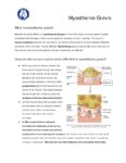 |
Myasthenia Gravis | NANOS | This is an autoimmune condition where the body's immune system has damaged receptors on your muscles and can result in double vision or drooping lid. | Myasthenia Gravis; Patient Brochure |
| 114 |
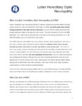 |
Hereditary Optic Neuropathy (Leber's Hereditary Optic Neuropathy) | NANOS | Hereditary Optic Neuropathy - A hereditary optic neuropathy is caused by a genetic variant (or mutation) that causes dysfunction of the neurons (nerve cells) which form the optic nerve. The optic nerve sends information from the back of the eye to the vision center in the brain.The two most common t... | Hereditary Optic Neuropathy; Patient Brochure |
| 115 |
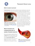 |
Transient Vision Loss | NANOS | Vision loss that is temporary (transient) is a common problem and has many potential causes.Patients with temporary vision loss often do not have any abnormalities on their eye examination, especially once the vision has returned to normal. | Transient Vision Loss; Patient Brochure |
| 116 |
 |
Cogan Lid Twitch | Hari Anandarajah, BA | A 50-year-old woman presented with ptosis of her left eyelid for 6 months. Several exam findings including variable and fatigable ptosis, and Cogan lid twitch, raised suspicion for Myasthenia Gravis. Acetylcholine receptor binding, blocking, and modulating antibodies were negative, and single fiber ... | Lid Twitch; Myasthenia Gravis |
| 117 |
 |
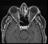
Indirect Carotid Cavernous Fistula | Edsel Ing, MD, PhD, FRCSC | A 67-year-old woman had delayed initial diagnosis of her right low flow carotid cavernous fistula (CCF) during the coronavirus disease (COVID-19) pandemic due to difficulty detecting ocular signs via online virtual examinations. Her right eye conjunctival erythema and proptosis with medial rectus en... | Carotid Cavernous Fistula; Misdiagnosis; Radiology |
| 118 |
 |
Giant Cell Arteritis: Diagnostic Prediction Models, Temporal Artery Biopsy and Epidemiology | Edsel Ing MD, PhD FRCSC MPH CPH MIAD MEd MBA, | Giant cell arteritis (GCA) is the most common primary vasculitis in the elderly and can cause irreversible blindness, aortitis, and stroke. Diagnostic confirmation of GCA usually entails temporal artery biopsy (TABx) - a time-consuming and invasive test, or ultrasound. The primary treatment of GCA i... | Giant Cell Arteritis; Diagnostic Prediction Model; Epidemiology; Temporal Artery Biopsy; Differential Diagnosis |
| 119 |
 |
Radiation Optic Neuropathy | Khawla Elnour; Amanda Henderson, MD | A video describing optic neuropathy related to radiation. | Radiation; Neuropathy |
| 120 |
 |
Vogt-Koyanagi-Harada Syndrome | Shwetha Mudalegundi, Medical Student; Amanda D. Henderson, MD | Vogt-Koyanagi-Harada (VKH) syndrome is a rare disorder that affects several body systems. Here we take a broad look at the presentation and pathophysiology of VKH, with a more specific focus on the relevant eye findings. Since much is not known about VKH, we explore the current standards for diagnos... | Vogt-Koyanagi-Harada Syndrome; Uveitis |
| 121 |
 |
Surgical Management of Strabismus | Michelle S. Attzs, MBBS, FRCOphth | This is a brief introduction to the surgical management of strabismus. It includes the key elements of the work up for a patient about to undergo strabismus surgery, introduces the basics on surgical techniques including adjustable sutures, and discusses the complications associated with this surger... | Strabismus; Surgery; Ocular Motility; Adjustable Sutures; Esotropia; Exotropia; Complications |
| 122 |
 |
Direct-Indirect Ophthalmoscopy (DIO) | Irina Krikova, PA-C; Eric Caskey, MD; Alison Crum, MD; Kathleen Digre, MD; James Gilman, CRA, FOPS; Levi Goldfarb, MBA, MD Candidate; Bradley Katz, MD; Ethan Peterson, Videographer; Meagan Seay, DO; Judith Warner, MD | A slideshow describing the use of the direct ophthalmoscope. | Ophthalmoscopy |
| 123 |
 |
John Cunningham Virus | Alison Gibbons; Amanda D. Henderson, MD | This learning object is a narrated Power Point presentation describing the features of, risk factors for, and clinical presentations of the John Cunningham, or JC, virus. It includes a discussion of various immunosuppressed states, including HIV, use of natalizumab (a disease-modifying therapy that ... | John Cunningham Virus; JC Virus; Natalizumab |
| 124 |
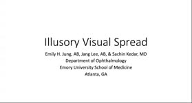 |
Illusory Visual Spread | Emily H. Jung, AB; Jang Lee, AB; Sachin Kedar, MD | Illusory visual spread is a form of visual perseveration resulting in images appearing to spread beyond it normal confines. In this video we describe a patient with Parkinson's disease and dementia, who developed various forms of visual hallucination including palinopsia and illusory visual spread. ... | Visual Perseveration; Illusory Visual Spread; Visual Hallucination |
| 125 |
 |
Corectopia | Meagan Seay, DO | These are photos of a patient with unilateral corectopia. This patient's corectopia is of unclear etiology and possibly related to birth trauma. | Corectopia; Unilateral; Photos |