Collection of materials relating to neuro-ophthalmology as part of the Neuro-Ophthalmology Virtual Education Library.
NOVEL: https://novel.utah.edu/
TO
- NOVEL233
| Title | Creator | Description | Subject | ||
|---|---|---|---|---|---|
| 201 |
 |
Hypothalamus: Neuroanatomy Video Lab - Brain Dissections | Suzanne S. Stensaas, PhD | Gross specimens are used to demonstrate the area of the hypothalamus and its relationship to surrounding structures. Both endocrine and autonomic functions are explored using diagrams. Mention is made of the direct hypothalamic response to circulating hormones and other substances such as sodium. Th... | Hypothalamus; Brain; Dissection |
| 202 |
 |
Three Critical Vertical Pathways: Neuroanatomy Video Lab - Brain Dissections | Suzanne S. Stensaas, PhD | There is one motor and two sensory pathways that must be mastered. Pain and temperature from the body travel together and vibration and proprioception travel in another pathway each reaching perception in the cortex. Voluntary motor control starts in the cerebral cortex and connects with a motor neu... | Spinothalamic Tract; Dorsal Column-Medical Lemniscus Pathway; Posterior Column; Vertical Pathway; Brain; Dissection |
| 203 |
 |
The Spinal Cord & Monosynaptic Reflex: Neuroanatomy Video Lab - Brain Dissections | Suzanne S. Stensaas, PhD | The spinal cord's relationship to the foramina, discs and spinal nerves is demonstrated on a model. The dura, ganglia and rootlets are shown as well as the gray and white matter in gross sections at different levels. A model of the cord is used to demonstrate and describe the anatomy of a monosynapt... | Spinal Cord; Monosynaptic Reflex; Brain; Dissection |
| 204 |
 |
Limbic System: Neuroanatomy Video Lab - Brain Dissections | Suzanne S. Stensaas, PhD | The decision was made to present a simplified description of a much more complex system using animations to construct a 3D image. Papez circuit is shown on gross specimens with mention of its involvement in memory. The role of the amygdala in fear and the olfactory cortex in temporal lobe epilepsy a... | Limbic System; Hippocampus; Brain; Dissection |
| 205 |
 |
The Most Important Pathway: Motor Control: Neuroanatomy Video Lab - Brain Dissections | Suzanne S. Stensaas, PhD | The origin of the corticospinal tract in the cerebral cortex is traced through gross sections of the hemisphere and brain stem to the spinal cord. Using an animation, the terms upper and lower motor neuron are defined and clinical signs and symptom listed. | Corticospinal Tract; Cerebral Cortex; Motor Neuron; Brain; Dissection |
| 206 |
 |
The Unfixed Spinal Cord: Neuroanatomy Video Lab - Brain Dissections | Suzanne S. Stensaas, PhD | The spinal cord's relationship to the foramina, discs and spinal nerves is demonstrated on a model. The dura, ganglia and rootlets are shown as well as the gray and white matter in gross sections at different levels. A model of the cord is used to demonstrate and describe the anatomy of a monosynapt... | Unfixed Spinal Cord; Spinal Cord; Brain; Dissection |
| 207 |
 |
Olfactory System: Neuroanatomy Video Lab - Brain Dissections | Suzanne S. Stensaas, PhD | Beginning with the location of the sensory cells within the skull the axons are traced into the cranial cavity. Demonstration of the olfactory bulb, olfactory tract and it termination in the forebrain and temporal lobe are indicated. Trauma and meningiomas can produce loss of small (anosmia). Degene... | Olfactory System; Olfactory Bulb; Anosmia; Brain; Dissection |
| 208 |
 |
The Meninges: Neuroanatomy Video Lab - Brain Dissections | Suzanne S. Stensaas, PhD | The epidural, subdural and subarachnoid spaces are demonstrated and discussed with respect to trauma and disease. The relationship of the brainstem and cerebellum to the tentorium demonstrates the vulnerability of the brain stem to increased supratentorial pressure and herniation. Arachnoid granulat... | Meninges; Brain; Dissection |
| 209 |
 |
Orientation: The Planes of the Brain: Neuroanatomy Video Lab - Brain Dissections | Suzanne S. Stensaas, PhD | Terms such as anterior, posterior, inferior and superior are introduced with respect to the hemispheres as well as the brain stem. Terms such as rostral and caudal or dorsal and ventral can mean different things in different areas. Sections in three planes (frontal, axial, and sagittal) are demonstr... | Frontal; Axial; Sagittal; Brain; Dissection |
| 210 |
 |
The Visual Pathway: Neuroanatomy Video Lab - Brain Dissections | Suzanne S. Stensaas, PhD | A brief review of the anatomy of the eye and the photic stimulation of the receptors is followed by a gross exploration of the visual pathway from the optic nerve, chiasm, and tract to the thalamus stressing how the left part of the visual world reaches the right hemisphere. Visual fields are relate... | Visual Pathway; Brain; Dissections |
| 211 |
 |
The Normal Unfixed Brain: Neuroanatomy Video Lab - Brain Dissections | Suzanne S. Stensaas, PhD | The consistency and vulnerability of the brain is demonstrated along with the clear and glistening pia and arachnoid and the tough dura. The cushioning function of the CSF is stressed and the features are pointed out on the ventral surface. The uncus and temporal lobes are normal with arteries free ... | Brain; Dissections |
| 212 |
 |
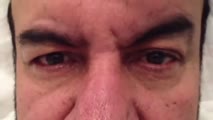
Cogan's Lid Twitch Sign | Raed Behbehani, MD | Cogan's lid twitch sign is a twitch sign of he upper lid upon looking straight from a sustained downgaze position. It is associated with Ocular Myasthenia Gavis. | Myasthenia; Ptosis; Lid Twitch |
| 213 |
 |
Upbeat Nystagmus | Raed Behbehani, MD, | A patient with a brain stem syndrome due to demyelination and upbeat nystagmus. | Upbeat Nystagmus |
| 214 |
 |
Downbeat Nystagmus Anti-GAD Cerebellar Syndrome | Raed Behbehani, MD | A patient with Anti-GAD positive Cerebellar syndrome with ataxia and opsoclonus due to downbeat nystagmus , treated with Baclofen with some improvement. | Downbeat Nystagmus |
| 215 |
 |
See-Saw Nystagmus | Raed Behbehani, MD | This nystagmus localizes to lesions supra/parasellar region (Large sellar and hypothalamic lesion) and is characterized by a see saw movement of elevation/intorsion of one eye and depression/extorsion of the other eye in a pendular fashion. This patient had a large pituitary macro-adenoma with supra... | See-Saw Nystagmus |
| 216 |
 |
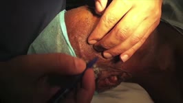
Optic Nerve Sheath Fenestration | Raed Behbehani, MD | Optic nerve sheath fenestration is performed to manage papilledema causing progressive loss of vision , due to raised intracranial pressure from Idiopathic Intracranial Hypertension or Cerebral Venous Sinus Thrombosis. The procedure is usually performed in cases of severe visual field loss or when m... | Optic Nerve Sheath Fenestration |
| 217 |
 |
Temporal Artery Biopsy | Raed Behbehani, MD | This is a video of Superficial Temporal Artery Biopsy done under local anaesthesia for a patient who was suspected to have Giant Cell Arteritis (GCA. GCA is vasculitis of the medium sized vessels than can lead to permanent visual loss by causing Arteritis Ischemic Optic Neuropathy. The diagnosis of ... | Temporal Artery Biopsy; Giant Cell Arteritis |
| 218 |
 |
Pulsating Exophthalmos | Raed Behbehani, MD | This patient had brain surgery with bone removal resulted in transmission of CSF pulsation into the orbit and pulsating exophthalmos. This sign can also be seen in patient with neurofibromatosis with hypoplasia of the sphenoid wing bone. | Pulsating Exophthalmos; Neurofibromatosis |
| 219 |
 |
Congenital Oculomotor Apraxia | Raed Behbehani, MD | Congenital Ocular Motor Apraxia is an uncommon condition that causes children to have difficulty moving their eyes horizontally or from side to side. They are usually unable to quickly move their eyes from side to side and often have to turn their head (head jerking) and not just their eyes to track... | Oculomotor Apraxia |
| 220 |
 |
Ocular Neuromyotonia | Raed Behbehani, MD | Ocular Neuromytonia is a characterised by by paroxysmal tonic contraction of the extraocular muscles supplied by the oculomotor nerve. It is has been reported after cranial radiation therapy, especially to the sellar-parasellar region and from compressive lesions such tumours or aneurysms. The patho... | Ocular Neuromyotania |
| 221 |
 |
Periodic Alternating Nystagmus | Raed Behbehani, MD | PAN is a nystagamus characterized by a cycle of uniderectional jerk nystagamus for 60-90 sec , a pause for 10-20 sec and a a cycle of a jerk nystagmus in the opposite direction for 60-90 sec. It is found in brain stem and cerebellar conditions as well as ocular albinism ( as in this patient). | Periodic Alternating Nystagmus |
| 222 |
 |
See-Saw Nystagmus | Raed Behbehani, MD | See-saw nystagmus is a localizing nystagmus to lesions of the sellar and parasellar region. "It's characterized by synchronous elevation and intorsion of one eye and depression and extorsion of the contra lateral eye . This patent has a craniopharyngioma, which was operated twice, optic atrophy and ... | See-Saw Nystagmus |
| 223 |
 |
Marcus Gunn Jaw Winking | Raed Behbehani, MD | Marcus Gunn Jaw Wink causes congenital ptosis and eyelid retraction associated with jaw movement or sucking. It's due to "miswiring" between 3rd and 5th cranial nerves. The treatment of ptosis in children is surgery to prevent amblyopia . | Jaw Winking; Marcus Gunn |
| 224 |
 |
Apraxia of Eyelid Opening | Raed Behbehani, MD | Patient has Parkinson disease and has developed this condition following deep brain stimulation. | Apraxia; Eyelid Opening |
| 225 |
 |
Pertinent Pupillary Problems | Karl C. Golnik, MD | Pupil Exam is a narrated PowerPoint that covers the basic of examining pupils. | Pupil Exam; Pupil |