Best known for his world-renowned neuro-ophthalmology unit based at the University of California, San Francisco, William Hoyt, MD collected here more than 850 of his best images covering a wide range of disorders.
William F. Hoyt, MD, Professor Emeritus of Ophthalmology, Neurology and Neurosurgery, Department of Ophthalmology, University of California, San Francisco.
NOVEL: https://novel.utah.edu/
TO
1 - 25 of 9
| Title | Description | Type | ||
|---|---|---|---|---|
| 1 |
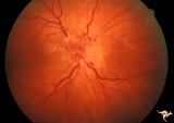 |
B404 Disc Swelling, Radiation Papillopathy | Marked vascular changes in the swollen optic disc. Probably not blind. Male. Right eye. Anatomy: Optic disc. Pathology: Axoplasmic stasis due to ischemia. Disease/ Diagnosis: Radiation Papillopathy; Optic neuropathy. Clinical: Visual loss after radiation therapy. | Image |
| 2 |
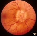 |
Bilateral Papilledema | Right eye. Bilateral Papilledema from vitamin A toxicity. Vitamin A pseudotumor cerebri syndrome in a 25 year old weight lifter. Anatomy: Optic disc. Pathology: Bilateral papilledema. Disease/Diagnosis: Pseudotumor due to vitamin A toxicity and weight lifting. Clinical notes: Headache, weight lifter... | Image |
| 3 |
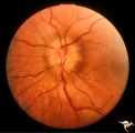 |
Bilateral Papilledema | Left eye. Bilateral Papilledema from vitamin A toxicity. Vitamin A pseudotumor cerebri syndrome in a 25 year old weight lifter. Anatomy: Optic disc. Pathology: Bilateral papilledema. Disease/Diagnosis: Pseudotumor due to vitamin A toxicity and weight lifting. Clinical notes: Headache, weight lifter. | Image |
| 4 |
 |
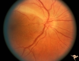
C110 Papillitis, Retrobulbar Neuritis | AIDs papillitis. Segmental. Note inflammatory focus on temporal side of disc. 29 year old homosexual male. Visual field shows huge blind spot. Anatomy: Optic disc. Pathology: Axoplasmic stasis due to inflammation. Disease/ Diagnosis: AIDS papillitis / AIDS Optic neuritis. Clinical: Visual symptoms d... | Image |
| 5 |
 |
C112 Papillitis, Retrobulbar Neuritis | Woman with herpes. Acute retinal necrosis with papillitis and arcuate neuro-retinitis. Right eye. Pair with C1_13. Reference: Margolis T, Irvine AR, Hoyt WF, Hyman R. Acute retinal necrosis syndrome presenting with papillitis and arcuate neuroretinitis. Ophthalmology. 1988 Jul;95(7):937-40. Anatomy:... | Image |
| 6 |
 |
C113 Papillitis, Retrobulbar Neuritis | Woman with herpes. Acute retinal necrosis with papillitis an arcuate neuro-retinitis. Right eye. Notice the large arcuate defect extending fromt he disc to the retina of retinal necrosis. Pair with C1_12. Anatomy: Optic disc; Retina. Pathology: Axoplasmic stasis due to inflammation; Retinal necrosis... | Image |
| 7 |
 |
F203 Optic Nerve Sheath Meningioma | Optic nerve sheath meningioma. Note optociliary vessels on the disc. The disc is partially atrophic and blurred by previous edema. The cause of the choroidal scar was not determined. Anatomy: Optic disc. Pathology: Chronic optic disc swelling caused by optic nerve sheath meningioma. DIsease/ Diagnos... | Image |
| 8 |
 |
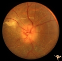
H40 Segmental Hypoplasia, Retinal, Tilted (Dysverted) Disc | 60 year old woman with incidental bitemporal visual field depression. Extreme tilting of optic disc with inferior nasal segmental hypoplasia. Nasal retinal ectasia. Same patient as H_41. Anatomy: Optic disc; retina. Pathology: Hypoplasia secondary to retinal lesion. Disease/ Diagnosis: Segmental opt... | Image |
| 9 |
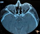 |
H41 Segmental Hypoplasia, Retinal, Tilted (Dysverted) Disc | CT scan of patient in H_40 showing marked nasal ectasia of the eyeballs. CT scan shows obliquely inserted optic nerves and marked nasal dysplasia of the eyeballs. Anatomy: Optic disc; retina. Pathology: Hypoplasia secondary to retinal lesion. Disease/ Diagnosis: Segmental optic disc hypoplasia. Imag... | Image |
1 - 25 of 9