Collection of materials relating to neuro-ophthalmology as part of the Neuro-Ophthalmology Virtual Education Library.
NOVEL: https://novel.utah.edu/
TO
- NOVEL728
| Title | Creator | Description | Subject | ||
|---|---|---|---|---|---|
| 551 |
 |
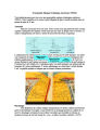
Thyroid Eye Disease (Simplified Chinese) | NANOS | This is an autoimmune condition where your body's immune system is producing factors that stimulate enlargement of the muscles that move the eye. | Thyroid Eye Disease; Thyroid Orbitopathy; Patient Brochure |
| 552 |
 |
Thyroid Eye Disease (Traditional Chinese) | NANOS | This is an autoimmune condition where your body's immune system is producing factors that stimulate enlargement of the muscles that move the eye. | Thyroid Eye Disease; Thyroid Orbitopathy; Patient Brochure |
| 553 |
 |
Anisocoria (German) | NANOS | Pupil in the right eye and left eye are not the same size. | Anisocoria; Patient Brochure |
| 554 |
 |
Anterior Ischemic Optic Neuropathy (German) | NANOS | Loss of blood supply to the optic nerve results in diminished visual acuity. | Anterior Ischemic Optic Neuropathy; Patient Brochure |
| 555 |
 |
Blepharospam (German) | NANOS | Uncontrolled blinking, squeezing, and eyelid closure that occurs in both eyes without an apparent environmental cause. | Blepharospasm; Patient Brochure |
| 556 |
 |
Hemifacial Spasm (German) | NANOS | Involuntary contractions, called "spasms," of the muscles on one side of the face. The affected side of the face seems to "scrunch up" while the other side of the face remains normal. | Hemifacial Spasm; Patient Brochure |
| 557 |
 |
Homonymous Hemianopia (German) | NANOS | This refers to an absence of vision towards one side of the visual world in each eye. The damage that caused this problem is in the brain and not in the eyes. | Homonymous Hemianopia; Patient Brochure |
| 558 |
 |
Microvascular Cranial Nerve Palsy (German) | NANOS | Microvascular cranial nerve palsy is one of the most common causes of double vision in the older poulation. They are often referred to as "diabetic" palsies. They will resolve without leaving any double vision. | Microvascular Cranial Nerve Palsy; Patient Brochure |
| 559 |
 |
Pituitary Tumor (German) | NANOS | Pituitary tumors are benign (non-cancerous) overgrowth of cells that make up the pituitary gland (the master gland that regulates other glands in the body). | Pituitary Tumor; Patient Brochure |
| 560 |
 |
Pseudotumor Cerebri (German) | NANOS | This is a condition in which high pressure inside your head can cause problems with vision and headache. | Pseudotumor Cerebri; Patient Brochure |
| 561 |
 |
Anisocoria (French) | NANOS | Pupil in the right eye and left eye are not the same size. | Anisocoria; Patient Brochure |
| 562 |
 |
Anterior Ischemic Optic Neuropathy (French) | NANOS | Loss of blood supply to the optic nerve results in diminished visual acuity. | Anterior Ischemic Optic Neuropathy; Patient Brochure |
| 563 |
 |
Blepharospasm (French) | NANOS | Uncontrolled blinking, squeezing, and eyelid closure that occurs in both eyes without an apparent environmental cause. | Blepharospasm; Patient Brochure |
| 564 |
 |
Homonymous Hemianopia (French) | NANOS | This refers to an absence of vision towards one side of the visual world in each eye. The damage that caused this problem is in the brain and not in the eyes. | Homonymous Hemianopia; Patient Brochure |
| 565 |
 |
Microvascular Cranial Nerve Palsy (French) | NANOS | Microvascular cranial nerve palsy is one of the most common causes of double vision in the older poulation. They are often referred to as "diabetic" palsies. They will resolve without leaving any double vision. | Microvascular Cranial Nerve Palsy; Patient Brochure |
| 566 |
 |
Optic Neuritis (French) | NANOS | In the most common form of optic neuritis, the optic nerve has been attacked by the body's overactive immune system and results in decreased vision. | Optic Neuritis; Patient Brochure |
| 567 |
 |
Pseudotumor Cerebri (French) | NANOS | This is a condition in which high pressure inside your head can cause problems with vision and headache. | Pseudotumor Cerebri; Patient Brochure |
| 568 |
 |
Genetic Information Nondiscrimination Act of 2008 | Senate and House of Representatives of the United States of America in Congress | Law to prohibit discrimination on the basis of genetic information with respect to health insurance and employment. | Genetics |
| 569 |
 |
Optic Disc Drusen (German) | NANOS | Optic disc drusen are abnormal deposits of protein-like material in the optic disc - the front part of the optic nerve. | Optic Disc Drusen; Patient Brochure |
| 570 |
 |
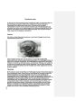
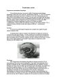
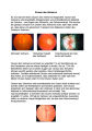
Central/Branch Retinal Artery Occlusion (PowerPoint) | AAO/NANOS - American Academy of Ophthalmology / North American Neuro-Ophthalmology Society | Occlusion of a branch or central retinal artery may result in acute visual loss. The ophthalmoscopic findings are retinal whitening due to ischemic retina in the distribution of the occluded artery. Sparing or selective involvement of cilioretinal artery branches may occur. Patients with a central r... | Central/Branch Retinal Artery Occlusion |
| 571 |
 |
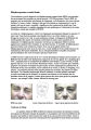
Chest CT Thymoma (Guest Lecture) | Robert A. Novelline, MD | The patient is a 46 year old woman who presented in July 1977 with horizontal double vision lasting two weeks. Three weeks later the left upper eyelid started to droop and by the end of the day the eye was closed. She had no ptosis of the right eye and no generalized fatigue. She consulted an intern... | Unilateral Ptosis; Unilateral Lid Retraction; Myasthenic Lid Twitch; External Ophthalmoplegia; Ocular Myasthenia Gravis; Tensilon Test; Thymolipoma; Generalized Myasthenia Gravis; Unilateral Myasthenia Gravis; Myasthenic Ptosis; Lid Retraction; Lid Twitch |
| 572 |
 |
Chiasmal Herniation (PowerPoint) | AAO/NANOS - American Academy of Ophthalmology / North American Neuro-Ophthalmology Society | This woman was 61 years old when she underwent initial neuro-ophthalmologic evaluation. Twenty-three years earlier, she had undergone removal of a pituitary adenoma followed by radiation therapy. At that time, she had noted a preoperative visual field defect that improved somewhat after the surgery.... | Chiasmal Herniation |
| 573 |
 |
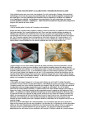
Evolution of Optociliary Veins with Perioptic Nerve Sheath | William F. Hoyt, PhD | Series of images showing progression of disc swelling and macular degeneration. Pathology: Optociliary Vein. Disease/Diagnosis: Perioptic nerve sheath meningioma evolution. Clinical notes: Visual Loss. | Optic Disc Atrophy with Special Features; Optociliary Veins; Shunt Vessels (Meningioma) |
| 574 |
 |
Exposed Drusen (PowerPoint) | William F. Hoyt, PhD | PP25a: Left eye: Severe visual field defect. PP25b: right eye with exposed drusen and field loss: visual field defects; PP25c: right eye visual field PP25d: left eye visual field. | Pseudopapilledema; Exposed Drusen |
| 575 |
 |
Brain MRI in Multiple Sclerosis (Guest Lecture) | Anne G. Osborn, MD | The patient is a 25 year old woman who was in excellent health until 4 days prior to admission when she noted blurred vision and horizontal double vision on lateral gaze to right and left. Past History: Negative for strabismus as a child. No previous episodes of transient neurological symptoms. Fami... | Bilateral Internuclear Ophthalmoplegia; Abducting Nystagmus; Normal Convergence; Gaze Evoked Upbeat Nystagmus; Gaze Evoked Downbeat Nystagmus; Saccadic Dysmetria; Multiple Sclerosis; Horizontal Saccadic Dysmetria |