Best known for his world-renowned neuro-ophthalmology unit based at the University of California, San Francisco, William Hoyt, MD collected here more than 850 of his best images covering a wide range of disorders.
William F. Hoyt, MD, Professor Emeritus of Ophthalmology, Neurology and Neurosurgery, Department of Ophthalmology, University of California, San Francisco.
NOVEL: https://novel.utah.edu/
TO
Filters: Date: "1970" Collection: "ehsl_novel_wfh"
| Title | Description | Type | ||
|---|---|---|---|---|
| 26 |
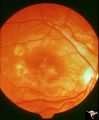 |
Retinal (Macular) Involvement in Subacute Sclerosing Pan Encephalopathy | Retinal (macular) involvement in Subacute Sclerosing Pan Encephalopathy (SSPE). Chronic macular changes with bilateral blindness. Anatomy: Retina. Pathology: Cerebral and retinal degeneration. Disease/Diagnosis: Subacute Sclerosing Pan Encephalopathy (SSPE). Clinical: Progressive visual loss and pro... | Image |
| 27 |
 |
Retinal Signs of Atheromatous Embolization | Retinal signs of atheromatous embolization. Central retinal artery occlusion by soft atheromatous debris (mostly fibrin) causing blindness. Anatomy: Retina. Pathology: Carotid atheromatous disease. Disease/Diagnosis: Carotid atheromatous vascular disease. Clinical: Blindness. | Image |
| 28 |
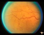 |
Slow Flow (Chronic Hypoxic) Retinopathy | Examples of Slow flow (chronic hypoxic) retinopathy showing dilated and tortuous retinal veins and multiple capillary hemorrhages. This kind of retinopathy is produced by impaired arteriole circulation to the retina from various causes. Anatomy: Retina. Pathology: Ophthalmic artery venous malformati... | Image |
| 29 |
 |
Slow Flow (Chronic Hypoxic) Retinopathy | Examples of Slow flow (chronic hypoxic) retinopathy showing dilated and tortuous retinal veins and multiple capillary hemorrhages. This kind of retinopathy is produced by impaired arteriole circulation to the retina from various causes. Anatomy: Retina. Pathology: Ophthalmic artery venous malformati... | Image |
| 30 |
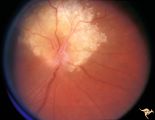 |
Tuberous Sclerosis | Tuberous Sclerosis. Astrocytic hamartoma of the optic disc. Anatomy: Optic disc. Pathology: Astrocytic hamartoma. Disease/Diagnosis: Tuberous sclerosis. Clinical: No visual symptoms. | Image |
| 31 |
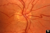 |
Unilateral Buried Drusen | PP20a: Right eye. Normal disc without optic cup.PP20b: Left eye. Buried drusen nasally and exposed drusen at the temporal margin. Boy. Anatomy: Optic disc. Pathology: Drusen of the optic disc. Disease/Diagnosis: Drusen of the optic disc. Clinical: Normally functioning eye with drusen. Right eye nor... | Image |
| 32 |
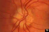 |
Unilateral Buried Drusen | PP20a: Right eye. Normal disc without optic cup.PP20b: Left eye. Buried drusen nasally and exposed drusen at the temporal margin. Boy. Anatomy: Optic disc. Pathology: Drusen of the optic disc.. Disease/Diagnosis: Drusen of the optic disc.. Clinical: Normally functioning eye with drusen. Right eye n... | Image |
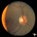
| 33 |
 |
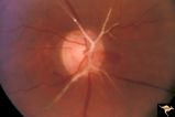
Unilateral Papilledema | Left eye. Patient had tumor on right side. Right sided large meningioma. optociliary shunt at 10:00. Foster Kennedy. Anatomy: Optic disc. Pathology: Unilateral papilledema. Disease/Diagnosis: Meningioma of the brain. Clinical: Headache. | Image |
| 34 |
 |
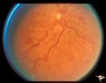
Unilateral Papilledema | Right eye. Patient had tumor on right side. Right sided large meningioma. Disc edema due to tumor. 29 year old black woman. The right disc has mild temporal pallor. Anatomy: Optic disc. Pathology: Uninaleral papilledema. Disease/Diagnosis: Meningioma of the brain. Clinical: Headache. | Image |
| 35 |
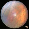 |
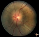
Von Hippel Lindau Disease | Von Hippel Lindau Disease. Retinal photograph showing small whitish hemangiomablastoma. Note the dilated arterial and venous channels entering and leaving the tumor. Anatomy: Retina. Pathology: Hemangioblastoma. Disease/Diagnosis: Von Hippel Lindau disease. Clinical: No visual symptoms. | Image |