Best known for his world-renowned neuro-ophthalmology unit based at the University of California, San Francisco, William Hoyt, MD collected here more than 850 of his best images covering a wide range of disorders.
William F. Hoyt, MD, Professor Emeritus of Ophthalmology, Neurology and Neurosurgery, Department of Ophthalmology, University of California, San Francisco.
NOVEL: https://novel.utah.edu/
TO
Filters: Date: "1969" Collection: "ehsl_novel_wfh"
| Title | Description | Type | ||
|---|---|---|---|---|
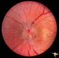
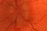
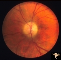
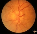
| 1 |
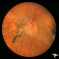 | A301a Disc Swelling, Chorioretinal Disease | a and b same eye. Bad chorioretinal scars with disc swelling. Anatomy: Optic disc. Pathology: Unknown. | Image |
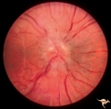
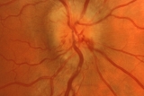
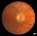
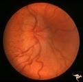
| 2 |
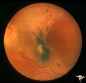 | A302b Disc Swelling, Chorioretinal Disease | Bad chorioretinal scars with disc swelling. Temporal extent of chorioretinal scarring. A and B are the same eye. Anatomy: Optic disc. Pathology: Unknown. | Image |
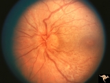
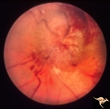
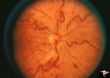
| 3 |
 | B101 Disc Swelling, Ischemic Papillopathies, AION | Pallid swelling in course of acute AION. 48 year old man who developed disc swelling after a flu like illness, then developed AION. Anatomy: Optic disc. Pathology: Axoplasmic stasis due to ischemia. Disease/ Diagnosis: AION. Clinical: Visual loss after flu-like illness. | Image |
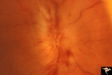
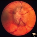
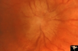
| 4 |
 | B203 Disc Swelling, Diabetic Papillopathy | Disc swelling in a diabetic woman. Recovered without visual loss. Right eye. Pair with B2_04. Anatomy: Optic disc. Pathology: Axoplasmic stasis due to ischemia. Disease/ Diagnosis: Diabetic papillopathy. Clinical: Visual loss with recovery. | Image |
| 5 |
 | B204 Disc Swelling, Diabetic Papillopathy | Disc swelling in a diabetic. Recovered without visual loss. Left eye. Pair with B2_03. Anatomy: Optic disc. Pathology: Axoplasmic stasis due to ischemia. Disease/ Diagnosis: Diabetic papillopathy. Clinical: Visual loss with recovery. | Image |
| 6 |
 | B403 Disc Swelling, Radiation Papillopathy | Man with blind eye. Ischemic hemorrhages. Vitreous haze. Anatomy: Optic disc. Pathology: Axoplasmic stasis due to ischemia. Disease/ Diagnosis: Radiation papillopathy; optic neuropathy. Clinical: Visual loss after radiation therapy. | Image |
| 7 |
 | Bilateral Papilledema | Left eye. Has intra-retinal exudate and unusual vascular changes in the optic disc. Pre-pubertal girl. Anatomy: Optic disc. Pathology: Bilateral papilledema; exudative deposits in macula. Disease/Diagnosis: Pseudotumor. Clinical: Pubertal girl; headaches. | Image |
| 8 |
 | Bilateral Papilledema | Right eye. Pre-pubertal girl. Anatomy: Optic disc. Pathology: Bilateral papilledema; exudative deposits in macula. Disease/Diagnosis: Pseudotumor. Clinical notes: Pubertal girl; headaches. | Image |
| 9 |
 | Bilateral Papilledema from Occipital Tumor | Left eye. Bilateral hemorrhagic papilledema. Occipital glioma. Woman. Anatomy: Optic disc. Pathology: Papilledema. Disease/Diagnosis: Hemorrhagic papilledema from occipital glioma. | Image |
| 10 |
 | Bilateral Papilledema from Occipital Tumor | Right eye. Bilateral hemorrhagic papilledema. Occipital glioma. Right hemianopia. Woman. Anatomy: Optic disc. Pathology: Papilledema. Disease/Diagnosis: Hemorrhagic papilledema from occipital glioma. | Image |
| 11 |
 | Bilateral Papilledema from Pseudotumor | Left eye. Atrophic changes in left optic disc. Chronic papilledema with involution to atrophy on the left. Woman. Anatomy: Optic disc. Pathology: Bilateal papilledema; atrophic papilledema. Disease/Diagnosis: Pseudotumor. Clinical: Headache. | Image |
| 12 |
 | Bilateral Papilledema from Pseudotumor | Right eye. Chronic papilledema. Woman. Anatomy: Optic disc. Pathology: Bilateral papilledema; atrophic papilledema. Disease/Diagnosis: Pseudotumor. Clinical notes: Headache. | Image |
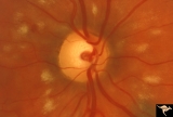
| 13 |
 | C04 Pits of the Optic Disc | Right eye. Man. Large temporal pit. Macular detachment. Anatomy: Optic disc. | Image |
| 14 |
 | C05 Pits of the Optic Disc | Right eye. Pigmented pit. Woman. Anatomy: Optic disc. | Image |
| 15 |
 | C101 Papillitis, Retrobulbar Neuritis | Resolved. Associated polycythemia. Papillitis after flu in patient with polycythemia. Homosexual male. Anatomy: Optic disc. Pathology: Axoplasmic stasis due to inflammation. Disease/ Diagnosis: Post infectious papillitis. Clinical: Visual loss after the flu.. | Image |
| 16 |
 | C102 Papillitis, Retrobulbar Neuritis | Inflammatory papillitis in 25 year old woman. Resolved completely. Anatomy: Optic disc. Pathology: Axoplasmic stasis due to inflammation. Disease/ Diagnosis: Inflammatory optic papillitis. Clinical: Visual loss. | Image |
| 17 |
 | Chronic Papilledema due to Brain Tumor | Right eye. Chronic papilledema wth white centrally located exudates in a man with hemispheric glioma. Anatomy: Optic disc. Pathology: Papilledema. Disease/Diagnosis: Chronic papilledema. | Image |
| 18 |
 | Chronic Papilledema due to Brain Tumor | Left eye. Chronic papilledema with white centrally located exudates in a man with hemispheric glioma. Anatomy: Optic disc. Pathology: Papilledema. Disease/Diagnosis: Chronic papilledema. | Image |
| 19 |
 | Chronic Papilledema due to Brain Tumor - Resolved | Left eye - same as P_40b - follow up after 4 months. Chronic papilledema resolved after treatment showing residual atrophy. | Image |
| 20 |
 | Chronic Papilledema due to Brain Tumor - Resolved | Right eye - same as P_40a - follow up after 4 months. Chronic papilledema resolved after treatment showing residual atrophy. Anatomy: Optic disc. Pathology: Papilledema. Disease/Diagnosis: Chronic papilledema. | Image |
| 21 |
 | E03 Disc Swelling with Central Retinal Vein Occlusion | 36 year old woman with visual obscurations of right eye. Early CRVO, papillophlebitis. Steroid responsive. Anatomy: Optic disc; Retina. Pathology: Central retinal vein occlusion. Disease/ Diagnosis: Disc swelling due to central retinal vein occlusion. Clinical: Decreased vision in right eye. Acuity ... | Image |
| 22 |
 | F106 Histiocytosis Infiltrate of Disc | More fully developed and chronic histiocytosis infiltrate of right disc with simultaneous infiltration of the hypothalamus with skin lesions on eye lids and chest. Same patient as F1_05, one year later. Anatomy: Optic disc. Pathology: Histiocytosis infiltrate of disc. Disease/ Diagnosis: Histiocytos... | Image |
| 23 |
 | F201 Optic Nerve Sheath Meningioma | Right eye. Woman with ophthalmoplegia proptosis for 14 years. Visual field reduced due to optic nerve sheath meningioma. Notice large optociliary vessel temporally. Anatomy: Optic disc. Pathology: Chronic optic disc swelling caused by optic nerve sheath meningioma. Disease/ Diagnosis: Chronic optic ... | Image |
| 24 |
 | F2b05 Optic Disc Swelling from Optic Glioma | Optic disc swelling from optic glioma. Patient had Neurofibromatosis (NF1). Left eye. 7 year old girl. 20/100 acuity. Glioma of the left optic nerve. Anatomy: Optic disc. Pathology: Optic nerve glioma. Disease/ Diagnosis: Optic nerve swelling secondary to retrobulbar optic glioma | Image |
| 25 |
 | G204 Purtchers Traumatic Retinopathy | Right eye. Blind due to chest crush with broken ribs. 18 year old male. Anatomy: Optic disc. Pathology: Varied peripapillary ischemic retinopathy. Disease/ Diagnosis: Purtchers traumatic retinopathy. | Image |