Best known for his world-renowned neuro-ophthalmology unit based at the University of California, San Francisco, William Hoyt, MD collected here more than 850 of his best images covering a wide range of disorders.
William F. Hoyt, MD, Professor Emeritus of Ophthalmology, Neurology and Neurosurgery, Department of Ophthalmology, University of California, San Francisco.
NOVEL: https://novel.utah.edu/
TO
1 - 25 of 7
| Title | Description | Type | ||
|---|---|---|---|---|
| 1 |
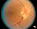 |
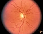
B201 Disc Swelling, Diabetic Papillopathy | Bilateral simultaneous diabetic papillopathy with marked exudation and remarkable recovery of vision. Right eye. Pair with B2_2b. Anatomy: Optic disc. Pathology: Axoplasmic stasis due to ischemia. Disease/ Diagnosis: Diabetic papillopathy. Clinical: Visual loss. | Image |
| 2 |
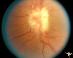 |
B202 Disc Swelling, Diabetic Papillopathy | Bilateral diabetic papillopathy with marked exudation and remarkable recovery of vision. Left eye. Pair with B2_1a. Anatomy: Optic disc. Pathology: Axoplasmic stasis due to ischemia. Disease/ Diagnosis: Diabetic papillopathy. Clinical: Visual loss with recovery. | Image |
| 3 |
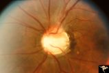 |
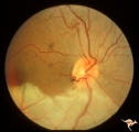
C08 Pits of the Optic Disc | Left eye. Large cavitary anomaly (pit). Man. 20/100 visual acuity. Superior nasal visual field defect. May not have a central retinal artery. Anatomy: Optic disc. Clinical: Man. 20/100 visual acuity. Superior nasal visual field defect. | Image |
| 4 |
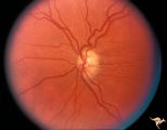 |
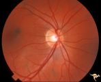
H52 Superior Segmental Optic Hypoplasia (SSOH) Topless Disc Syndrome | High exit point of central retinal vessels. Superior choroidal crescent. Complete loss of nerve fiber layer entering disc from above. Inferior altitudinal field defect. Type 1 diabetic mother. Anatomy: Optic disc. Pathology: Superior segmental optic hypoplasia (SSOH). Disease/ Diagnosis: Congenital ... | Image |
| 5 |
 |
H76 Superior Segmental Optic Hypoplasia (SSOH) Topless Disc Syndrome | SSOH. Right eye. Anatomy: Optic disc. Pathology: Superior segmental optic hypoplasia (SSOH). Disease/ Diagnosis: Congenital anomaly. | Image |
| 6 |
 |
Neurofibromatosis-1 | Normal appearing optic disc with dark pigmented choroidal nevi. The patient had NF-1 and had a subclinical optic glioma on the left eye. This is the right eye. Anatomy: Optic disc. Pathology: Choroidal nevus. Disease/Diagnosis: Neurofibromatosis type 1. Clinical: No visual symptoms. | Image |
| 7 |
 |
Vascular Disc Anomalies - Prepapillary Arterial Loop | Complication of prepapillary arterial loop causing occlusion of the inferior retinal arterial and resulting inferior retinal infarction. Appears to be a black thrombus in the apex of the arterial loop. Anatomy: Optic disc. Pathology: Arterial loop with retinal artery infarction. Disease/Diagnosis: B... | Image |
1 - 25 of 7