Best known for his world-renowned neuro-ophthalmology unit based at the University of California, San Francisco, William Hoyt, MD collected here more than 850 of his best images covering a wide range of disorders.
William F. Hoyt, MD, Professor Emeritus of Ophthalmology, Neurology and Neurosurgery, Department of Ophthalmology, University of California, San Francisco.
NOVEL: https://novel.utah.edu/
TO
1 - 25 of 14
| Title | Description | Type | ||
|---|---|---|---|---|
| 1 |
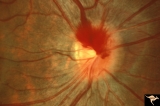 |
A406 Disc Swelling, Vitreous Effects | Prepapillary hemorrhage. Partial posterior vitreous detachment in myopic Asian patient. Reference: Katz B, Hoyt WF. Intrapapillary and peripapillary hemorrhage in young patients with incomplete posterior vitreous detachment. Signs of vitreopapillary traction. Ophthalmology. 1995 Feb;102(2):349-54. A... | Image |
| 2 |
 |
A408 Disc Swelling, Vitreous Effects | Prepapillary hemorrhage. Partial posterior vitreous detachment in myopic Asian patient. Reference: Katz B, Hoyt WF. Intrapapillary and peripapillary hemorrhage in young patients with incomplete posterior vitreous detachment. Signs of vitreopapillary traction. Ophthalmology. 1995 Feb;102(2):349-54. A... | Image |
| 3 |
 |
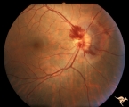
Buried Drusen with Sub-retinal Neovascular Net | Buried drusen with sub-retinal neovascular net. Both PP29a and PP29b are left eye: 17 year old girl; Visual acuity 10/400. Anatomy: Optic disc. Pathology: Drusen of the optic disc. Disease/Diagnosis: Drusen of the optic disc. Clinical notes: Loss of central vision due to subretinal neovascularizatio... | Image |
| 4 |
 |
Buried Drusen with Sub-retinal Neovascular Net | Buried drusen with sub-retinal neovascular net. This is the same left eye. Appearance of the central retina of the left eye. Both PP29a & b are left eye: 17 year old girl; Visual acuity 10/400. Anatomy: Optic disc. Pathology: Drusen of the optic disc. DIsease/Diagnosis: Drusen of the optic disc. Cl... | Image |
| 5 |
 |
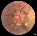
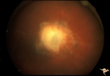
C105 Disc Edema with Systemic Lupus | Mild disc edema blurs the inferior disc margin. Flourescein angiogram in D1_06. Same patient as D1_06 an D1_07. Man. Anatomy: Optic disc. Pathology: Axoplasmic stasis due to vasculitis (Lupus). Disease/ Diagnosis: Lupus papillopathy. | Image |
| 6 |
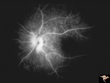 |
D106 Disc Edema with Systemic Lupus | Flourescein angiogram shows evidence of vascular papillopathy. (Lupus) Same patient as D1_05 an D1_07. Anatomy: Optic disc. Pathology: Axoplasmic stasis due to vasculitis (Lupus). Disease/ Diagnosis: Lupus papillopathy. | Image |
| 7 |
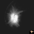 |
D107 Disc Edema with Systemic Lupus | Late stage Flourescein angiogram showing flourescein leakage on the disc and around the neighboring vessels. Note this amount of edema could not be appreciated in the colored fundus image D1_05. Same patient as D1_06 an D1_05. Anatomy: Optic disc. Pathology: Axoplasmic stasis due to vasculitis (Lupu... | Image |
| 8 |
 |
Drusen with Horizontal Retinal Folds | PP35a: Right eye. Buried drusen. PP35b: Left eye. Buried drusen with retinal folds. 21 year old woman. Anatomy: Optic disc. Pathology: Drusen of the optic disc. Disease/Diagnosis: Drusen of the optic disc. | Image |
| 9 |
 |
Drusen with Horizontal Retinal Folds | PP35: Right eye. Buried drusen. PP35b: Left eye. Buried drusen with retinal folds. 21 year old woman. Anatomy: Optic disc. Pathology: Drusen of the optic disc. Disease/Diagnosis: Drusen of the optic disc. | Image |
| 10 |
 |
G101 Evulsion | BB injury of the optic nerve with traumatic evulsion. The missile went through the eyeball and hit the optic disc. Anatomy: Optic disc. Pathology: Optic nerve has been evulsed. Disease/ Diagnosis: Evulsion of the optic disc. | Image |
| 11 |
 |
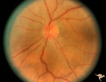
Neurofibromatosis-2 | This is the ocular fundus in a patient with NF-2 showing a preretinal membrane that extends from the temporal disc margin toward the macula. The optic disc shows low grade papilledema caused by one of the patient's acoustic neurinomas. The membrane has caused horizontal folds on the retinal surface.... | Image |
| 12 |
 |
Neurofibromatosis-2 | CPERH (choroidal pigment epithelial retinal hamartoma) lesion in a patient with NF-2. Note the oblique superficial retinal traction folds running toward the center of the main lesion. 51 year old man. Anatomy: Retina. Pathology: Hamartoma. Disease/Diagnosis: Neurofibromatosis type 2. Clinical: Fiel... | Image |
| 13 |
 |
Venous Anomalies - Exit Anomalies | Choriovaginal vein. Choroid in myopic disc is raining into optic disc. Choriovaginal vein entering disc ege at 9:00. Kraupa type 2. Peripapillary atrophy in highly myopic eye. Same patient, magnified, as V_38. Anatomy: Optic disc. Pathology: Congenital anomaly of choroidal venous drainage. Disease/D... | Image |
| 14 |
 |
Venous Anomalies - Exit Anomalies | Choriovaginal vein. Choroid in myopic disc is draining into optic disc. Choriovaginal vein entering disc ege at 9:00. Kraupa type 2.Peripapillary atrophy in highly myopic eye. Same patient as V_39. Anatomy: Optic disc. Pathology: Congenital anomaly of choroidal venous drainage. Disease/Diagnosis: C... | Image |
1 - 25 of 14