Best known for his world-renowned neuro-ophthalmology unit based at the University of California, San Francisco, William Hoyt, MD collected here more than 850 of his best images covering a wide range of disorders.
William F. Hoyt, MD, Professor Emeritus of Ophthalmology, Neurology and Neurosurgery, Department of Ophthalmology, University of California, San Francisco.
NOVEL: https://novel.utah.edu/
TO
Filters: Collection: "ehsl_novel_wfh"
| Title | Description | Type | ||
|---|---|---|---|---|
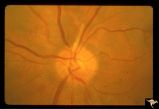
| 651 |
 | Resolution of Papilledema Following Optic Nerve Sheath Decompression (ONSD) | Left eye. 17 year old boy. Cryptococcal meningitis. Resolution of papilledema following optic nerve sheath fenestration (ONSF) on November 1, 1974. Same eye as P_53a on November 7, 1974, one week following ONSF. Anatomy: Optic disc. Pathology: Papilledema. Disease/Diagnosis: Resolving papilledema. | Image |
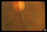
| 652 |
 | Resolution of Papilledema Following Optic Nerve Sheath Decompression (ONSD) | Left eye. 17 year old boy. Cryptococcal meningitis. Same eye as P_53a. Increased papilledema. August 1974. Anatomy: Optic disc. Pathology: Papilledema. Disease/Diagnosis: Resolving papilledema. | Image |
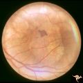
| 653 |
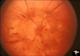
 | Retinal (Macular) Involvement in Subacute Sclerosing Pan Encephalopathy | Retinal (macular) involvement in Subacute Sclerosing Pan Encephalopathy (SSPE). Chronic macular changes with bilateral blindness. Anatomy: Retina. Pathology: Cerebral and retinal degeneration. Disease/Diagnosis: Subacute Sclerosing Pan Encephalopathy (SSPE). Clinical: Progressive visual loss and pro... | Image |
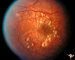
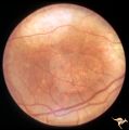
| 654 |
 | Retinal (Macular) Involvement in Subacute Sclerosing Pan Encephalopathy | Retinal (macular) involvement in Subacute Sclerosing Pan Encephalopathy (SSPE). Acute macular changes with bilateral blindness. Anatomy: Retina. Pathology: Cerebral and retinal degeneration. Disease/Diagnosis: Subacute Sclerosing Pan Encephalopathy (SSPE). Clinical: Progressive visual loss and progr... | Image |
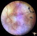
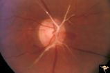
| 655 |
 | Retinal (Macular) Involvement in Subacute Sclerosing Pan Encephalopathy | Retinal (macular) involvement in Subacute Sclerosing Pan Encephalopathy (SSPE). Note optic disc pallor. Anatomy: Retina. Pathology: Cerebral and retinal degeneration. Disease/Diagnosis: Subacute Sclerosing Pan Encephalopathy (SSPE). Clinical: Progressive visual loss and progressive cerebral degenera... | Image |
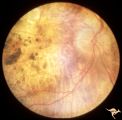
| 656 |
 | Retinal (Macular) Involvement in Subacute Sclerosing Pan Encephalopathy | Retinal (macular) involvement in Subacute Sclerosing Pan Encephalopathy (SSPE). Note interesting microvascular changes associated with the retinal disease. Anatomy: Retina. Pathology: Cerebral and retinal degeneration. Disease/Diagnosis: Subacute Sclerosing Pan Encephalopathy (SSPE). Clinical: Progr... | Image |
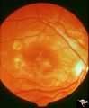
| 657 |
 | Retinal Degeneration Associated with Spastic Paraplegia | Retinal pigmentary degeneration concentrated around the optic discs in a patient with spastic paraplegia. Inverted retinitis pigmentosa where bone spicules are concentrated around the disc and maculae instead of the periphery. Left eye. Anatomy: Retina. Pathology: Cerebellar spinal degenerative dise... | Image |
| 658 |
 | Retinal Degeneration Associated with Spastic Paraplegia | Retinal pigmentary degeneration concentrated around the optic discs in a patient with spastic paraplegia. Inverted retinitis pigmentosa where bone spicules are concentrated around the disc and maculae instead of the periphery. Right eye. Anatomy: Retina. Pathology: Cerebellar spinal degenerative dis... | Image |
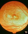
| 659 |
 | Retinal Pigmentary Degeneration with Progressive External Ophthalmoplegia | This 55 year old woman has pigmentary retinal degeneration with progressive external ophthalmoplegia (PEO). (Kearns-Sayre Syndrome). Anatomy: Retina. Pathology: Mitochondrial disease. Disease/Diagnosis: Progressive external ophthalmoplegia (PEO). Clinical: Can't move eyes. | Image |
| 660 |
 | Retinal Pigmentary Degeneration with Progressive External Ophthalmoplegia | This 55 year old woman has pigmentary retinal degeneration with progressive external ophthalmoplegia (PEO). (Kearns-Sayre Syndrome). Anatomy: Retina. Pathology: Mitochondrial disease. Disease/Diagnosis: Progressive external ophthalmoplegia (PEO). Clinical: Can't move eyes. | Image |
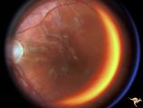
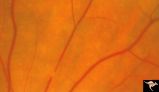
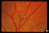
| 661 |
 | Retinal Signs of Atheromatous Embolization | Retinal signs of atheromatous embolization. Central retinal artery occlusion by soft atheromatous debris (mostly fibrin) causing blindness. Anatomy: Retina. Pathology: Carotid atheromatous disease. Disease/Diagnosis: Carotid atheromatous vascular disease. Clinical: Blindness. | Image |
| 662 |
 | Retinal Signs of Atheromatous Embolization | Retinal signs of atheromatous embolization. Series shows event in progress. R3_A19c shows embolus has reached the bifurcation and a second embolus (below) is beginning its transit along the same path. Anatomy: Retina. Pathology: Carotid atheromatous disease. Disease/Diagnosis: Carotid atheromatous v... | Image |
| 663 |
 | Retinal Signs of Atheromatous Embolization | Retinal signs of atheromatous embolization. Series shows event in progress. R3_A19d shows progress along the same transit path for both emboli. Anatomy: Retina. Pathology: Carotid atheromatous disease. Disease/Diagnosis: Carotid atheromatous vascular disease. Clinical: No visual symptom. | Image |
| 664 |
 | Retinal Signs of Atheromatous Embolization | Retinal signs of atheromatous embolization. Series shows event in progress. R3_A19a shows atheromatous embolus traveling up the arteriole. Anatomy: Retina. Pathology: Carotid atheromatous disease. Disease/Diagnosis: Carotid atheromatous vascular disease. Clinical: No visual symptom. | Image |
| 665 |
 | Retinal Signs of Atheromatous Embolization | Retinal signs of atheromatous embolization. Series shows event in progress. R3_A19b shows embolus approaching the bifurcation. Anatomy: Retina. Pathology: Carotid atheromatous disease. Disease/Diagnosis: Carotid atheromatous vascular disease. Clinical: No visual symptom. | Image |
| 666 |
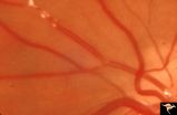 | Retinal Signs of Atheromatous Embolization | Retinal signs of atheromatous embolization. Note shiny cholesterol plaques in retinal arterial. Anatomy: Retina. Pathology: Intraluminal cholesterol crystals. Disease/Diagnosis: Carotid atheromatous vascular disease. Clinical: No visual symptoms. | Image |
| 667 |
 | Retinal Signs of Atheromatous Embolization | Retinal signs of atheromatous embolization. Embolic occlusion of central retinal artery by white thrombis (probably fibrin.) Patient had a myocardial infarction. Anatomy: Retina. Pathology: Occlusion of a central retinal artery. Disease/Diagnosis: Carotid atheromatous vascular disease. Clinical: Sud... | Image |
| 668 |
 | Retinal Signs of Atheromatous Embolization | Retinal signs of atheromatous embolization. White changes in the arteriolar wall are called plasma bleeding. They are produced by scratches in the endothelium from cholesterol embolization. Anatomy: Retina. Pathology: Plasma bleeding, Post cholesterol embolization. Disease/Diagnosis: Carotid atherom... | Image |
| 669 |
 | Retinal Signs of Atheromatous Embolization | Retinal signs of atheromatous embolization. Note the way the cholesterol emboli stick at arteriole bifurcation. Note second plaque hidden at the juncture below. Anatomy: Retina. Pathology: Intraluminal cholesterol crystals. Disease/Diagnosis: Carotid atheromatous vascular disease. Clinical: No visua... | Image |
| 670 |
 | Retinal Signs of Atheromatous Embolization | Retinal signs of atheromatous embolization. Note shiny cholesterol plaques in retinal arterial. Anatomy: Retina. Pathology: Occlusion of the superior retinal arteriole at the level of the optic disc. Disease/Diagnosis: Carotid atheromatous vascular disease. Clinical: Inferior visual field loss from ... | Image |
| 671 |
 | Retinal Signs of Atheromatous Embolization | Retinal signs of atheromatous embolization. Example of plasma bleeding in the arteriole wall secondary to cholesterol embolization. Anatomy: Retina. Pathology: Focal capillary bleeding. Disease/Diagnosis: Carotid atheromatous vascular disease. Clinical: Patient had several attacks of amaurosis fugax... | Image |
| 672 |
 | Retinal Signs of Atheromatous Embolization | Retinal signs of atheromatous embolization. Atheromatous embolism in retinal arteriole branch with associated minimal opacification. Anatomy: Retina. Pathology: Carotid atheromatous disease. Disease/Diagnosis: Carotid atheromatous vascular disease. Clinical: Sudden inferior visual field loss. | Image |
| 673 |
 | Retinal Signs of Atheromatous Embolization | Retinal signs of atheromatous embolization. Atheromatous embolism in a patient who suffered sudden visual loss in his left eye. Anatomy: Retina. Pathology: Post cholesterol embolization. Disease/Diagnosis: Carotid atheromatous vascular disease. Clinical: Sudden visual loss in left eye. | Image |
| 674 |
 | Retinal Signs of Atheromatous Embolization | Retinal signs of atheromatous embolization. Cholesterol embolization causing capillary bleeding. Anatomy: Retina. Pathology: Focal capillary bleeding. Disease/Diagnosis: Carotid atheromatous vascular disease. Clinical: No visual symptoms. | Image |
| 675 |
 | Retinal Signs of Atheromatous Embolization | Retinal signs of atheromatous embolization. Documentation of atheromatous embolus appearing at bifurcation during photographic session. R3_A18a shows no embolus and b shows new embolus. Anatomy: Retina. Pathology: Carotid atheromatous disease. Disease/Diagnosis: Carotid atheromatous vascular disease... | Image |