| OCR Text |
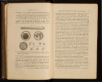
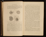
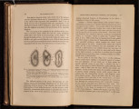
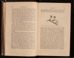
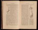
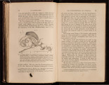
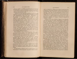
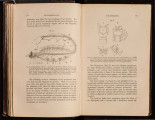
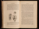
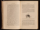
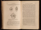
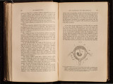
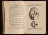
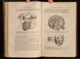
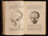
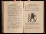
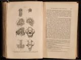
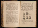
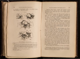
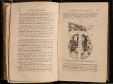
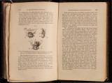
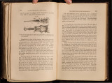
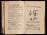
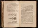
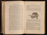
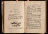
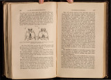
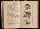
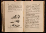
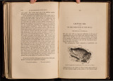
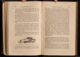
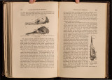
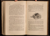
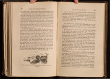
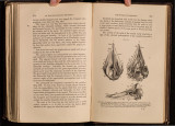
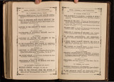
Show 154 ON THE STRUCTURE OF THE SKULL. the tyn1panic, is J{ercluingius' ossicle, " vi.,,r aciculm Inajoris caput adrequans." (Fig. 61, B, Op.O.) Fig. Gl. Fig. 61.- Development of the temporal bone. A, from a footus 5i inches long, showing tile commencing pro-oti c and opisth otic ossification. B, from a f<l'tus 8! inch es long. The ossification in the tegmen tympani is no longer visible fi·om without, but its continuation backwards over the l:>Uperi or, and part. of ~ h e posterior, Yertical semicircular canal is visible behind the squamosal. The epioti.c oss~ fi cation has made its nppearance, and the hinder extremity of the opisthoti(: ossificatiOn appears behind the tympanic as the "third o:ssicle" of Ker ckringi us. U, from a fretus l Ot inches long, the " tria ossicula " beginning to unite into the pars mastoidea. D, from a footus 10! inches long, t.he tria ossicula anchylosed. F.R., foramen rot?tndum. S.sc, superior semicircular canal. THE DEVELOPMENT OF THE HUMAN SKULL. 155 Last.ly, the thirrl ossicle, "scutum ovale referens," is that developed upon the posterior part of the posterior vertical semicircular canal, which gives rise to the mastoid process. (Fig.· 61, B, Ep.O.) . Thus in a footus between the fifth and sixth months, the ' "pars mastoidea" exhibits the appearance represented in Fig. 61, B. Its upper part is cartilaginous, but its lower part is occupied by the three "ossicula" of Kerckringius, which have now come into contact, and begun to unite, though their primitive contours are perfectly distinct. The "pars mastoidea" of human anatomy is therefore not a single bone, but one, the "scutum ovale," combined with parts of two others; and as the "scutum ovale" is certainly the homologue of the bone I have termed Epiotic in the oviparous Vertebrata,* I propose to get rid of the confusing term "mastoid" altogether, and to call the specially "mastoid" part of the pars mastoidea, Epiotic. Of the three periotic bones thus developed, the pro-otic gives rise to most of the pars petrosa, which is visible in the interior of the skull (Fig. 59, A), investing, as it does, the roof of the cochlea, the superior, and part of the posterior, vertical semicircular canals, the internal auditory meatus, and forming the tegmen tympani. To it, in addition, is due the upper half of the circumference of the fenestra ovalis, and a considerable portion of the pars mastoidea, as has been stated above. The opisthotic bone constitutes all the pars petrosa visible on the base of the skull, furnishes the floor of the cochlea, surrounds the fenestra rotunda, and contributes half the contour of the fenestra ovalis; gives rise to the carotid canal by developing a lamella of bone, which gradually wraps itself round the carotid, and so converts the primitive groove for the vessel into a complete tube, at the same time furnishing the inner part of its floor to the tympanum. The lower edge of the squamosal is at first nearly straight, * Croonian Lecture. Proceedings of the Roy(J,l Society, 1858. In the absence of a sufficient knowledge of tho development of the human tempoml bone, I followed Hallmann in identifying the opisthotic of oviparous vertebrates with the mastoid of .Mammals nt the Lime tl1is lecture was delivered. |