| OCR Text |
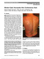
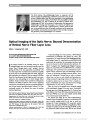
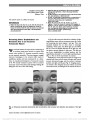
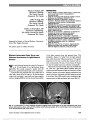
Show Midbrain Infarction Presenting With Monocular Elevation Palsy and Ptosis: Topographic Lesion Analysis Yun-Ju Choi, MD, Seung-Han Lee, MD, PhD, Man-Seok Park, MD, PhD, Byeong C. Kim, MD, PhD, Myeong-Kyu Kim, MD, PhD Abstract: A combination of monocular elevation palsy and ptosis is usually characteristic of an extra-axial lesion of the superior branch of the third nerve. We report an unusual case of monocular elevation palsy and ipsilateral ptosis due to midbrain infarction involving the third nerve fascicle. In addition, we conducted a review of the literature of similar cases and produced an overlay image of the magnetic resonance scans from these reports. The overlapping regions primarily were located in the midbrain between the red nucleus and cerebral peduncle. This correlated with involve-ment of the lateral portion of the third nerve fascicle containing fibers to the superior rectus and levator palpebrae. Journal of Neuro-Ophthalmology 2015;35:175-178 doi: 10.1097/WNO.0000000000000208 © 2015 by North American Neuro-Ophthalmology Society A partial third nerve palsy may due to involvement of the fascicular portion of the nerve within the brainstem (1,2). Reports following midbrain stroke include pupil-sparing third nerve palsy, isolated adduction palsy, isolated ptosis and mydri-asis, and monocular elevation palsy with ipsilateral ptosis (3-8). We describe a patient with findings mimicking a superior divi-sion third nerve palsy and review similar reported cases with associated magnetic resonance imaging (MRI) abnormalities. CASE REPORT A 75-year-old woman presented with a 2-day history of right ptosis and diplopia. She had a 13-year history of diabetes mellitus and hypertension. There was no ocular injection or pain, visual loss, or proptosis. Examination revealed complete right ptosis and limitation of supra-duction in the right eye with no abnormalities of adduction, infraduction, or pupillary reactivity (Fig. 1). The red glass test revealed vertical diplopia that increased in upward gaze and disappeared in downward gaze. Other cranial nerves, including the trigeminal and motor, sensory, and cerebellar systems were normal. Brain MRI did not reveal abnormal-ities in the right superior orbital fissure/cavernous sinus, orbit, or periorbital area. However, diffusion-weighted im-ages showed abnormal high-signal intensity in the right medial midbrain near the cerebral peduncle compatible with acute infarction (Fig. 2). Her symptoms gradually improved over 3 months. METHODS FOR LITERATURE REVIEW AND IMAGE OVERLAPPING We conducted a review of the literature of similar clinical manifestations and collected MRI scans of these cases. This was performed by a web-based search for both English (www. ncbi.nlm.nih.gov/pubmed) and Korean (www.koreamed.org) publications using the following terms: oculomotor nerve; third cranial nerve; stroke; hemorrhage; ischemia; infarct; elevation; paresis, palsy; midbrain; mesencephalon. Our search was conducted up to July, 2014. Articles were selected using predetermined criteria. These criteria excluded reports that lacked original patient data, did not provide description of ocular motor symptoms, and did not indicate MRI find-ings. We identified 30 articles, 23 of which were excluded due to the presence of only experimental data (n = 12) and other associated ophthalmologic abnormalities, such as oscil-lopsia and horizontal gaze palsy (n = 11). After a full-text review, we found 4 cases of monocular elevation palsy with ipsilateral ptosis in 3 (7-9). Four ar-ticles were excluded due to the lack of MR images. The demographics and clinical characteristics of the cases are shown in Table 1, and schematic diagrams of the MRI lesions appear in Figure 3. Department of Neurology (Y-JC, S-HL, M-SP, B-CK, M-KK), Chonnam National University Medical School, Gwangju, Korea; and Department of Neurology (Y-JC), Presbyterian Medical Center, Seonam University College of Medicine, Jeonju, Korea. The authors report no conflicts of interest. Address correspondence to Seung-Han Lee, MD, PhD, Department of Neurology, Chonnam National University Medical School, 8 Hak-dong, Dong-gu, Gwangju 501-757, Korea; E-mail: nrshlee@chonnam.ac.kr Choi et al: J Neuro-Ophthalmol 2015; 35: 175-178 175 Clinical Observation Copyright © North American Neuro-Ophthalmology Society. Unauthorized reproduction of this article is prohibited. FIG. 1. Nine cardinal positions of gaze reveal right ptosis and impaired elevation of the right eye. TABLE 1. Reports of superior division third nerve palsy due to midbrain stroke Author Age/Gender Other Neurologic Deficits MRI Findings Comment Celebisoy et al. (7) 62/F None Right ventral midbrain infarction (Fig. 3C) Park et al. (9) 57/F None Right ventral midbrain infarction (Fig. 3B) Park et al. (9) 71/F None Left ventral midbrain infarction (Fig. 3A)* Previous midbrain stroke involving inferior rectus and pupil Hriso et al. (8) 75/F Right hemiparesis; dysarthria Lesion involving cerebral peduncle and portion of midbrain tegmentum (image not shown) Current report 75/F None Right ventral midbrain infarction (Fig. 3D) *MRI scan reversed to create overlay schematic diagram (Fig. 3A) and MRI overlay image (Fig. 4). MRI, magnetic resonance imaging; M, male; F, female. FIG. 2. Axial (A), coronal (B), and sagittal (C) diffusion-weighted scans demonstrate a right paramedian midbrain infarction. 176 Choi et al: J Neuro-Ophthalmol 2015; 35: 175-178 Clinical Observation Copyright © North American Neuro-Ophthalmology Society. Unauthorized reproduction of this article is prohibited. We constructed an overlay image from each MRI scan containing the lesions using MRIcro software version 1.4 (www.mricro.com) (Fig. 4). The lesions of each patient were drawn manually onto transverse slices of the publicly available Montreal Neurological Institute brain, a T1- weighted template MRI scan, which is oriented to match the Talairach space. The template images containing the region of interest of each patient were summed up, and the overlapping area was shown in different colors accord-ing to the numbers of intersecting portions: 4 = red, 3 = green, 2 = blue, 1 = purple. One case with additional neurologic deficits due to a large midbrain stroke was excluded (8). The overlapping areas were primarily located in the midbrain between the red nucleus and cerebral peduncle. This area was correlated with previ-ously reported diagram of third nerve fascicular topogra-phy (1) (Fig. 5). DISCUSSION Our case highlights a rare manifestation of partial third nerve palsy characterized by monocular elevation palsy and ipsilateral ptosis due to midbrain infarction. Ischemia of the lateral portion of the fascicle that contains fibers innervating the superior rectus and levator palpebrae were affected in our patient. Several previous reports (7-10) also support our find-ings and are summarized in Table 1. Most cases had a focal ischemic lesion of the third nerve fascicle in the anterior portion of the midbrain between the cerebral peduncle and the red nucleus (Fig. 3). A case of lateral midbrain infarct with ptosis and upgaze palsy was documented in a patient with hemiparesis and dysarthria (8). In this patient, autopsy confirmed involvement of the lateral fascicular fibers. FIG. 3. Diagrams of 4 cases of ptosis and ipsilateral monocular elevation palsy due to midbrain infarction. A and B were derived from Ref. (9), (C) from Ref. (7), and (D) from the current report. FIG. 4. An overlay image created with MRIcro from MRI le-sions of the 4 patients with isolated monocular elevation palsy and ptosis. The area colored in red was most fre-quently involved (n = 4), followed by those colored in green (n = 3), blue (n = 2), and purple (n = 1). MRI, magnetic resonance imaging. Choi et al: J Neuro-Ophthalmol 2015; 35: 175-178 177 Clinical Observation Copyright © North American Neuro-Ophthalmology Society. Unauthorized reproduction of this article is prohibited. Most commonly, a superior division palsy of the third nerve is due to a lesion of the cavernous sinus/superior orbital fissure or orbit (3,8). Rarely, the cause may be an extra-axial ischemic neuropathy due to diabetes mellitus (6,11). In summary, a partial third nerve palsy in the absence of long tract signs can be caused by a fascicular brainstem lesion. Furthermore, midbrain infarction involving the lateral portion of the third nerve fascicle may present with isolated monocular elevation paresis and ipsilateral ptosis. REFERENCES 1. Ksiazek SM, Slamovits TL, Rosen CE, Burde RM, Parisi F. Fascicular arrangement in partial oculomotor paresis. Am J Ophthalmol. 1994;118:97-103. 2. Saeki N, Murai H, Mine S, Yamaura A. Fascicular arrangement within the oculomotor nerve MRI analysis of a midbrain infarct. J Clin Neurosci. 2000;7:268-270. 3. Breen LA, Hopf HC, Farris BK, Gutmann L. Pupil-sparing oculomotor nerve palsy due to midbrain infarction. Arch Neurol. 1991;48:105-106. 4. Chen L, Maclaurin W, Gerraty RP. Isolated unilateral ptosis and mydriasis from ventral midbrain infarction. J Neurol. 2009;256:1164-1165. 5. Ksiazek SM, Repka MX, Maguire A, Harbour RC, Savino PJ, Miller NR, Sergott RC, Bosley TM. Divisional oculomotor nerve paresis caused by intrinsic brainstem disease. Ann Neurol. 1989;26:714-718. 6. Massuci EF, Kurtzke JF. Diabetic superior branch palsy of the oculomotor nerve. Ann Neurol. 1980;7:493. 7. Celebisoy N, Celebisoy M, Tokucoglu F, Ozdemir I. Superior division paresis of the oculomotor nerve: report of four cases. Eur Neurol. 2006;56:50-53. 8. Hriso E, Masdeu JC, Miller A. Monocular elevation weakness and ptosis: an oculomotor fascicular syndrome? J Clin Neuroophthalmol. 1991;11:111-113. 9. Park JH, Jeong DS, Park SA, Lee TK, Sung KB. 3-D model of the oculomotor fascicular arrangement within the midbrain using brain MRI. Res Vestibul Sci. 2008;7:22-32. 10. Johnson LN, Castro O. Monocular elevation paresis and incomplete ptosis due to midbrain infarction involving the fascicular segment of the oculomotor nerve. J Clin Neuroophthalmol. 1992;12:73. 11. Collier J. Paralysis of the oculomotor nerve-trunks in diabetes. Proc R Soc Med. 1930;23:627. FIG. 5. Diagram of the topographic fascicular arrangement of the third nerve. The hatched area shows the area of involvement in our patient. IO, inferior oblique; IR, inferior rectus; LP, levator palpebrae; MR, medial rectus; P, constrictor pupillae; SR, superior rectus. 178 Choi et al: J Neuro-Ophthalmol 2015; 35: 175-178 Clinical Observation Copyright © North American Neuro-Ophthalmology Society. Unauthorized reproduction of this article is prohibited. |