| OCR Text |
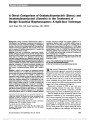
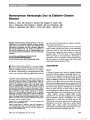
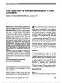
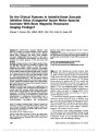
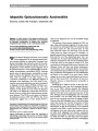
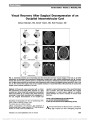
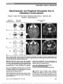
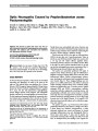
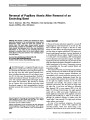
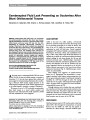
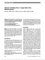
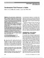
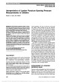
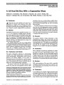
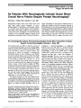
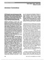
Show Optical Coherence Tomography in Papilledema: What Am I Missing? Randy Kardon, MD, PhD Background: Grading of papilledema severity is subjective and based on monocular fundus features of the optic nerve. Interobserver agreement on grading the severity of papillede-ma is poor among expert observers, even using well-defined criteria such as the Frisen scale, which is a non-continuous ordinal scale of grading. Furthermore, non-expert clinicians often find it difficult to properly view and interpret features of the optic nerve using ophthalmoscopy, which can lead to failure to diagnose papilledema in non-ophthalmologic care settings. This may delay treatment, which can result in vision loss. Distinguishing papilledema from pseudopa-pilledema can also be difficult when surface drusen are not easily identified. Once papilledema is diagnosed, it is often difficult to determine whether a reduction in optic nerve edema is due solely to improvement in the status of the nerve or whether this represents concomitant loss of axons and viable retinal ganglion cells, leading to a poor visual outcome. Timely advancement of treatment would occur if loss of neurons could be diagnosed at an earlier stage of evaluation while optic disc edema is still present. This review will critically assess the role of optical coher-ence tomography (OCT) in solving these problems by pro-viding an advanced imaging approach for diagnosis of papilledema and evaluating its severity on a continuous scale and evaluating the causes of visual loss in the set-ting of a swollen nerve. Methods Acquisition: The published literature (PubMed) was reviewed from 2000 to 2014 on the use of OCT for diagnosing papilledema, differentiating it from pseudopa-pilledema, providing a continuous scale of its severity and in evaluating causes of visual loss. Results: Recent evidence shows that OCT analysis of the retinal nerve fiber layer and retinal ganglion cell layer in papilledema can be associated with misleading artifacts due to layer segmentation failures. Newer 3D algorithms using neighboring locations help to overcome these problems. Disc volume appears to be a promising continuous measure of papilledema that is robust and has less associated artifacts. Buried optic disc drusen can be identified using enhanced depth OCT imaging, but recent studies have shown poor ability to differentiate papilledema from pseudopapilledema using OCT when the degree of disc evaluation is similar. Analysis of the retinal ganglion cell layer shows promise of early detection of vision loss due to neuronal injury. Subretinal fluid is easily identified with OCT and can help to identify a potentially reversible component of vision loss. Newer OCT imaging methods will allow the definition of capillaries and flow within them in and around the optic nerve head. Conclusions: Currently, the most useful OCT derived features relevant to papilledema are disc volume, subretinal fluid, buried disc drusen, and thickness of the retinal ganglion cell layer. Journal of Neuro-Ophthalmology 2014;34(Suppl):S10-S17 doi: 10.1097/WNO.0000000000000162 © 2014 by North American Neuro-Ophthalmology Society To the experienced neuro-ophthalmologist, the most relevant question is: Do we really need an imaging modality such as optical coherence tomography (OCT) to evaluate papilledema? Most clinicians feel that careful oph-thalmoscopy or digital fundus photography is more than adequate for diagnosing the presence of papilledema, deter-mining its severity and deciding on whether it has changed over time. After all, this is what has been done for years, so why do we need something more? The need for "something more" derives from a number of studies, including the multicenter National Institutes of Health sponsored Idiopathic Intracranial Pressure Treatment Trial: • Interobserver agreement on grading the severity of papilledema is poor among expert observers, even using well-defined criteria such as the Frisen scale, whether this is done using ophthalmoscopy or by grading of digital fundus photographs (1-3). • Non-expert clinicians often find it difficult to properly view the optic disc using ophthalmoscopy and to accu-rately interpret digital fundus photographs when using a non-mydriatic retinal camera. This can lead to failure to diagnose papilledema in non-ophthalmologic care settings such as emergency rooms, family practice Department of Ophthalmology and Visual Sciences, Neuro- Ophthalmology Division, University of Iowa, Hospitals and Clinics and Veterans Administration, Iowa City, Iowa. The author reports no conflicts of interest. Address correspondence to Randy Kardon, MD, PhD, Department of Ophthalmology (PFP), University of Iowa Hospital and Clinics, 200 Hawkins Drive, Iowa City, IA 52242; E-mail: randy-kardon@uiowa.edu S10 Kardon: J Neuro-Ophthalmol 2014; 34(Suppl): S10-S17 Original Contribution Copyright © North American Neuro-Ophthalmology Society. Unauthorized reproduction of this article is prohibited. offices, neurology and neurosurgery clinics, and may delay treatment, which can result in vision loss. • Distinguishing papilledema from pseudopapilledema is difficult when obvious surface drusen are not pres-ent. Buried drusen, when not calcified, may not be readily apparent using funduscopy, ultrasound, OCT, or computed tomographic (CT) scans. • It is often difficult to determine whether a reduction in optic disc edema is due solely to improvement in the status of the nerve, or whether this represents concomitant loss of axons and viable retinal ganglion cells, leading to a poor visual outcome. More timely advancement of treatment would occur if loss of neu-rons could be diagnosed at an earlier stage of evalua-tion while optic disc edema is still present. An important (and reachable) long-term goal is to provide a portable, low cost retinal imaging device with embedded software that would not require expertise for acquiring and making a diagnosis of papilledema or other optic nerve pathology. Ultimately, generation of an automated report providing diagnostic probability at the point of care and at the time of image acquisition is needed, which would bypass the need for a telemedicine reading center. Use of such a device would be adopted in clinical settings lacking easy access to ophthalmologists and neuro-ophthalmologists, such as in emergency rooms, family practice offices, neurology and neurosurgery clinics and inpatient units. This report will outline and define the critical need for new imaging modalities such as OCT and image analysis aimed at providing tools for improved diagnosis of papilledema, differentiating papillede-ma from pseudopapilledema and other causes of optic nerve edema, and for identifying early signs of retinal nerve loss to optimize treatment and prevent vision loss. DIAGNOSIS AND GRADING OF SEVERITY OF PAPILLEDEMA BASED ON FUNDUS FEATURES: ARE WE GOOD ENOUGH? Many clinicians are confident in their ability to accurately diagnose and grade the severity of papilledema, and most use the accepted standard of the Frisen grading scale (1). However, even in the original report by Frisen, there was intraobserver variability in grading of photographs on repeat testing, whether the grading was done by a medical student, resident, or expert (Fig. 1). Significant variability among experts in grading papilledema from digital fundus photo-graphs has also been demonstrated by Scott et al (2) (Fig. 2) and Sinclair et al (3) (Fig. 3). These studies give us pause in relying on humans (including experts) to accurately and reliably diagnose and grade papil-ledema. Efforts to refine and provide more specific criteria for each Frisen scale may help to improve reliability between observers. Since the Frisen scale consists of 6 grades (0-5) that are non-continuous, a further improvement would be to devise a continuous grading scale, based on structural features that could be objectively quantified by computerized image analysis of fundus images. Echegaray et al (4) have shown that quanti-tative analysis of digital fundus images is a promising approach and features that incorporate sharpness of the disc border, tex-ture of the retinal nerve fiber layer (RNFL), and discontinuity of blood vessels can be used by a machine classifier to assign a Frisen grade to a disc photograph. The next step would be to map features from digital fundus photography to a continuous scale based on OCT measurements of papilledema such as disc volume or thickness of the peripapillary retina (5,6). This would associate fundus photo features with OCT-based features, so that the quantification of papilledema on a continuous scale could be made (superseding the non-continuous Frisen scale) using either of these 2 imaging modalities. The imaging modal-ity to be used could be flexible and selected for a given patient based on availability and cost in a telemedicine setting where the patient enters the medical system. TOWARD A CONTINUOUS SCALE QUANTIFICATION OF PAPILLEDEMA SEVERITY (RETINAL NERVE FIBER LAYER, TOTAL RETINAL THICKNESS, AND DISC VOLUME) With the availability of time-domain OCT early in the 21st century, there had already been attempts at quantifying FIG. 1. Reproducibility for 3 different observers, who staged the same fundus photographs for disc swelling on 2 separate occasions. Each dot represents 1 photograph. The diagonals represent identity of stage numbers on test and retest. The first graph is for medical students, the second graph is for ophthalmology residents, and the third graph is for expert specialist neuro-ophthalmologists (1). Kardon: J Neuro-Ophthalmol 2014; 34(Suppl): S10-S17 S11 Original Contribution Copyright © North American Neuro-Ophthalmology Society. Unauthorized reproduction of this article is prohibited. papilledema using confocal microscopy with Heidelberg Retinal Tomography (HRT) and scanning laser polarimetry (SLP). HRT appeared promising (7) but was limited by the difficulty in defining an appropriate "reference plane" in the peripapillary retina for quantifying elevation of the nerve head above that plane, especially in higher grades of papil-ledema. Even in glaucomatous optic neuropathy, there was disagreement as to what portion of the retina was best suited for a reference plane that was not affected by the pathology being evaluated. Also, the availability of HRT was not wide-spread; only some academic institutions had the resources and interest to acquire the instrumentation for evaluating the optic nerve and retina using confocal laser microscopy. SLP, which also predated OCT, was gaining use for glau-coma evaluation and was based on changes in the retarda-tion of reflected polarized light from the RNFL that contained regularly oriented microtubules and microfila-ments, which could modify polarized light passing through it. Reports of its use showing thickening of the RNFL in papilledema were initially negative or only showed mild thickening (8,9); there was very little change in retardation of polarized light by axoplasmic flow stasis, since in most cases, the organization of the microfilament substructure was unaffected. However, SLP did reveal axon loss, similar to its use in glaucoma (8-10). Unlike HRT, OCT provided information on retinal thickening, and in particular, peripapillary thickening of the RNFL in papilledema without the need for a reference plane (11-21). Since OCT was based on actual thickness of the retinal layers, it complimented SLP, which primarily demonstrated loss of microtubule and microfilament orga-nization within the axon bundles. However, it was soon noticed that in the presence of moderate-to-severe papille-dema (Frisen Grade 3 or above), substantial thickening of the peripapillary RNFL would often cause the software algorithm that was used for determining the RNFL borders to fail in over one-third of the cases (2), causing inaccurate reporting of RNFL thickness. A significant improvement in the quantification of papilledema was achieved by segment-ing the total retinal thickness (TRT) in the same peripapil-lary scan, since the inner and outer borders of the retina can be more readily defined by automated software in the pres-ence of moderate to severe papilledema. The TRT was found to highly correlate with the RNFL thickness in eyes where the algorithm did not fail. Automated software segmentation of the retinal layers using a 3D graph-based approach has significantly improved the accuracy of defining the thickness of the retinal layers in FIG. 2. A. Four neuro-ophthalmology experts independently graded papilledema based on criteria outlined in the Frisen scale. B. Disagreement within on grade scale was common (43% of cases), and there were even cases where there were 2 or more grade scale difference between experts (3.5% of cases) at the higher grades (2). FIG. 3. Cross tabulation of the Frisen grades assigned to optic disc photographs by each pair of reviewers. Complete agreement between the reviewers is shaded in yellow. The red shading indicates disagreement between the reviewers in assigning a Frisen grade (3). S12 Kardon: J Neuro-Ophthalmol 2014; 34(Suppl): S10-S17 Original Contribution Copyright © North American Neuro-Ophthalmology Society. Unauthorized reproduction of this article is prohibited. papilledema (6), resulting in much fewer algorithm failures. With this approach, the entire surface of each retinal layer is determined within the scan volume using all of the surround-ing 3D features in the OCT scan and not just the features in each individual B scan. Wang et al (6) used this approach to segment the volume of the optic disc, which highly correlates with the RNFL, TRT, and Frisen grade of papilledema in patients with raised intracranial pressure (Fig. 4). ADVANCED FEATURE ANALYSIS OF THE DISC USING DIGITAL FUNDUS PHOTOGRAPHY AND OCT The ability to accurately derive an OCT-based, continuous measurement of papilledema (e.g., total disc volume, as explained in the previous section) provides an objective means of quantifying the severity of papilledema. The next step forward is to map other OCT and fundus based features to a continuous scale of disc volume, such as shape of the disc volume, deformation of Bruch's membrane at the neural canal and texture, along with fundus based features to further enhance the ability to differentiate papilledema from other forms of optic disc edema and pseudopapilledema. This will also allow quantification of features of digital fundus photo-graphs as shown in Figure 5 (e.g., obscuration of the disc margin, discontinuity of disc vessels, and texture of the peri-papillary nerve fiber layer) to be mapped to OCT disc vol-ume, and will provide the possibility of a continuous scale software measure of papilledema that can be derived and embedded in teleretinal imaging devices at the site of image capture for immediate diagnosis (4,6,22). Another OCT-based feature, which provides informa-tion about the direction of force vectors at the optic disc in papilledema, is the deformation of Bruch's membrane sur-rounding the neural canal due to a pressure differential between the retrobulbar optic nerve and vitreous cavity (Fig. 6). The shape characteristics of Bruch's membrane in this area, in terms of the degree of angling toward the vitreous, can help in monitoring of the force differential over time as the intracranial pressure changes and may also help to differentiate papilledema from other causes of optic disc edema or pseudopapilledema (23,24). This angle can vary to some degree within normal eyes without papille-dema. The angle may be slightly positive angling toward the vitreous, neutral and horizontal, or negative, angling toward the retrobulbar compartment. A very positive angle FIG. 4. A. Examples of how disc volume, derived from OCT, increases with increasing Frisen grade of papilledema (lower left section of figure). B scan sagittal sections are shown in the upper row and corresponding 3D disc volumes are shown in lower row. There is a highly linear correlation between Frisen grade and the disc volume (B) and disc volume and the retinal nerve fiber layer (RNFL) and total retinal thickness (C) (6). FIG. 5. Computerized image analysis features of fundus photos that are specific for papilledema and its degree of severity include, from left to right, texture ("entrophy") of the peripapillary retina (with insert), degree of definition of the disc border, vessel discontinuity index due to obscuration of retinal vessels by edematous overlying retinal nerve fiber layer, 3D disc volume derived from stereo pairs of disc photographs (4). Kardon: J Neuro-Ophthalmol 2014; 34(Suppl): S10-S17 S13 Original Contribution Copyright © North American Neuro-Ophthalmology Society. Unauthorized reproduction of this article is prohibited. in an eye suspected of having papilledema may be very helpful, but slightly positive or neutral angle does not rule out papilledema. A change in the angle from positive to less positive after treatment or after lumbar puncture would also enforce a suspicion of papilledema and would verify a treatment effect (Pat Sibony, MD, personal commu-nication, February 2014). Newer generation OCT instru-ments with enhanced depth penetration and longer wavelength light (exceeding 1 mm) provide even greater res-olution of deeper structures, such as Bruch's membrane, even in the presence of optic disc edema. DIFFERENTIATING PAPILLEDEMA FROM PSEUDOPAPILLEDEMA USING OCT The ability to differentiate papilledema due to raised intracranial pressure from other forms of optic disc edema or from pseudopapilledema can be challenging, particularly when the degree of edema is not severe (i.e., Frisen Grade 1 or 2). When calcified optic disc drusen are located superficially, the diagnosis is relatively easy and can be made with careful ophthalmoscopic observation. When calcified drusen are deep and buried under the surface, clinical observation may be equivocal, and the use of autofluores-cence, ultrasound, or observation of CT scans of the optic nerve have been useful. OCT has been used to differentiate papilledema from pseudopapilledema (25-33) (Fig. 7). Sometimes calcified drusen and their shadows, visualized on OCT, are not easy to distinguish from large, superficial blood vessels. Non-calcified drusen are not usually visualized, as they are presumed not to exhibit a significant difference in reflectivity from surrounding disc tissue. Often a patient with pseudopapilledema (with or without calcified drusen) may show visual field loss. In these eyes, the RNFL may appear thickened in some areas, presumably due to axoplasmic flow stasis, and thin in other areas, corresponding to locations of visual field loss. Another OCT approach to differentiating papilledema from pseudopapilledema is based on defining topographical shape characteristics of the elevated nerve head. In this approach, a machine classifier is used to define shape characteristics that are more likely to be associated with true papilledema and those characteristics that are more likely to be associated with pseudopapilledema. As outlined in the previous section, shape characteristics of Bruch's membrane may also help in differentiating papilledema from pseudopa-pilledema and other forms of optic disc edema not due to raised intracranial pressure. WHY IS MY PATIENT WITH PAPILLEDEMA LOSING VISION? DIFFERENTIATION OF VISUAL LOSS DUE TO OPTIC NEUROPATHY VS MACULOPATHY (FLUID AND SURFACE WRINKLING) When a patient with papilledema has best corrected vision of 20/25 or worse, then there is a concern for whether this may be caused by optic neuropathy, requiring more aggressive treatment, or whether it may be due to a macular abnormality such as subfoveal fluid or choroidal folds. The more benign retinal causes are relatively easy to diagnose with OCT and can help to resolve the uncertainty rather quickly. The most obvious sign that can be discerned with OCT is a neurosensory retinal detachment from peripa-pillary fluid between the retinal pigment epithelium and FIG. 6. A. Optic disc tissue in a patient with papilledema, showing the upward angling and displacement of Bruch's membrane (red arrows) in the right and left eyes. B. There is a change in displacement of Bruch's membrane in the same eye before and after treatment of raised intercranial pressure (23). S14 Kardon: J Neuro-Ophthalmol 2014; 34(Suppl): S10-S17 Original Contribution Copyright © North American Neuro-Ophthalmology Society. Unauthorized reproduction of this article is prohibited. photoreceptors that tracks into the fovea (34,35). On OCT B scans, the fluid appears dark with low reflectivity. The macular thickness is greater than surrounding areas without fluid on the color OCT thickness plot, and the probability plot shows the area with fluid to be signifi-cantly thicker than age matched normative data (Fig. 8). Decrease in vision due to fluid under the fovea is largely reversible and should not be considered a cause of vision loss requiring urgent management. However, rarely, with chronic papilledema, a subretinal neovascular membrane in the peripapillary retina may form and cause fluid that will not resolve unless treated more definitively with either intravitreal anti-vascular endothelial growth factor agents or laser treatment to the peripapillary area in the location of the membrane. Another retinal cause of decreased visual acuity is choroidal folds caused by distortion of the poste-rior globe by abnormal amounts of fluid under pressure in the subarachnoid space surrounding the optic nerve as it exits the globe. Choroidal folds can be recognized in OCT B scans, in the infrared fundus image, or on digital pho-tography and fundus examination. The folds may contrib-ute to metamorphopsia and are often reversible, but not always, with successful resolution of papilledema. Progres-sive optic neuropathy due to papilledema requires more aggressive, urgent treatment to attempt to minimize the degree functional and structural deficit and restore any reversible component of vision. A decline of visual acuity in the absence of macular fluid or folds is usually the most obvious sign of pro-gressive optic neuropathy in this setting. Since RNFL is thickened in papilledema, a reduction in its thickness, assessed by OCT, may be difficult to interpret and could represent either a reduction in disc edema due to improvement or due to axon loss (36-38). Kupersmith et al (10) have reported that axon loss in the presence of papilledema can be revealed by using SLP. Since SLP is sensitive to disorganization of axon microtubules and mi-crofilaments, which may be one of the earliest signs of axon disruption. However, this technology has become somewhat obsolete and was superseded by OCT for glau-coma diagnosis and monitoring. One next generation pro-totype OCT was developed with the capability of polarization assessment, but is not presently commercially available. As an alternative, assessment of ganglion cell loss by OCT in the setting of papilledema may be suitable for early detection of neuron loss in order to identify patients in need of more aggressive treatment. Since optic disc edema and axon swelling does not appear to directly affect the retinal ganglion cell layer thickness, allowing it to be an effective tool for the early diagnosis of progressive optic neuropathy. However, commercial algorithms for seg-menting the ganglion cell-inner plexiform layer (GCL-IPL) complex were designed for normal and glaucoma eyes and often fail in the presence of optic disc edema. OCT algorithms that take advantage of 3D information instead of just 2D information from single B scans are better suited to overcome this problem (6). The working assump-tion is that thinning of the GCL-IPL will reveal early signs of progressive optic neuropathy in the presence of papil-ledema. This will undoubtedly be the focus of studies in the near future to understand the usefulness of GCL-IPL thickness in the evaluation and monitoring of papilledema. FIG. 7. A very large, coalescing druse imaged in several SD-OCT modalities. A. Fundus photo with 2 vertical markers placed on either side of the druse (obtained with 3D disc scan on Topcon 3D OCT 2000). B. Low-resolution SD-OCT image, obtained on same 3D disc scan. C. High-resolution image, obtained with 7-line Raster on Topcon 3D OCT 2000. D. High-resolution (5-line Raster) image, obtained with Zeiss Cirrus HD-OCT (30). SD-OCT, spectral domain optical coherence tomography. Kardon: J Neuro-Ophthalmol 2014; 34(Suppl): S10-S17 S15 Original Contribution Copyright © North American Neuro-Ophthalmology Society. Unauthorized reproduction of this article is prohibited. Another recent development in OCT, which has possible relevance to understanding the pathogenesis of visual loss in papilledema due to ischemia relates to the visualization of optic nerve capillaries and capillary blood flow. Using phase contrast OCT, it is now possible to visualize capillaries and quantify flow within a capillary bed without the use of contrast agents (39). An example of OCT derived capillary flow in the normal and glaucomatous optic nerve head is shown in Figure 9. FIG. 8. Papilledema associated with a neurosensory retinal detachment between the left optic nerve and the fovea. A. The macula total retinal thickness plot shows the elevation in the area of the fluid (arrows). B. B scan through the detachment area reveals the fluid (dark reflective layer) between the pigment epithelium and the photoreceptors (arrow). C. 3D macula thickness plot demonstrates the elevation in the area of the detachment with fluid (arrow). FIG. 9. Disc photographs (A and C) and en face OCT angiograms (B and D) of the optic nerve head in representative normal (A and B) and PPG subjects (C and D). Both examples are from left eyes. In (B) and (D), the solid circles indicate the whole discs, and the dash circles indicate the temporal ellipses. A dense microvascular network was visible on the OCT angiography of the normal disc (B). This network was greatly attenuated in the glaucomatous disc (D) (39). OCT, optical coherence tomography; PPG, preperimetric glaucoma. S16 Kardon: J Neuro-Ophthalmol 2014; 34(Suppl): S10-S17 Original Contribution Copyright © North American Neuro-Ophthalmology Society. Unauthorized reproduction of this article is prohibited. REFERENCES 1. Frisen L. Swelling of the optic nerve head: a staging scheme. J Neurol Neurosurg Psychiatry. 1982;45:13-18. 2. Scott CJ, Kardon RH, Lee AG, Frisen L, Wall M. Diagnosis and grading of papilledema in patients with raised intracranial pressure using optical coherence tomography vs clinical expert assessment using a clinical staging scale. Arch Ophthalmol. 2010;128:705-711. 3. Sinclair AJ, Burdon MA, Nightingale PG, Mathews TD, Jacks A, Rating papilloedema: an evaluation of the Frisen classification in idiopathic intracranial hypertension. J Neurol. 2012;259:1406-1412. 4. Echegaray S, Zamora G, Yu H, Luo W, Soliz P, Kardon RH. Automated analysis of optic nerve images for detection and staging of papilledema. Invest Ophthalmol Vis Sci. 2011;52:7470-7478. 5. Tang L, Kardon RH, Wang JK, Garvin MK, Lee K. Quantitative evaluation of papilledema from stereoscopic color fundus photographs. Invest Ophthalmol Vis Sci. 2012;53:4490-4497. 6. Wang JK, Kardon RH, Kupersmith MJ, Garvin MK. Automated quantification of volumetric optic disc swelling in papilledema using spectral-domain optical coherence tomography. Invest Ophthalmol Vis Sci. 2012;53:4069-4075. 7. Trick GL, Vesti E, Tawansy K, Skarf B, Gartner J. Quantitative evaluation of papilledema in pseudotumor cerebri. Invest Ophthalmol Vis Sci. 1998;39:1964-1971. 8. Banks MC, Robe-Collignon NJ, Rizzo JF III, Pasquale LR. Scanning laser polarimetry of edematous and atrophic optic nerve heads. Arch Ophthalmol. 2003;121:484-490. 9. Laemmer R, Heckmann JG, Mardin CY, Schwab S, Laemmer AB. Detection of nerve fiber atrophy in apparently effectively treated papilledema in idiopathic intracranial hypertension. Graefes Arch Clin Exp Ophthalmol. 2010;248:1787-1793. 10. Kupersmith MJ, Kardon R, Durbin M, Horne M, Schulman J. Scanning laser polarimetry reveals status of RNFL integrity in eyes with optic nerve head swelling by OCT. Invest Ophthalmol Vis Sci. 2012;53:1962-1970. 11. Savini G, Barboni P, Carbonelli M, Carelli V, Sadun AA. Optical coherence tomography for optic disc edema. Arch Ophthalmol. 2011;129:1245-1246;author reply 1246-1247. 12. Savini G, Bellusci C, Carbonelli M, Zanini M, Carelli V, Sadun AA, Barboni P. Detection and quantification of retinal nerve fiber layer thickness in optic disc edema using stratus OCT. Arch Ophthalmol. 2006;124:1111-1117. 13. Ophir A, Karatas M, Ramirez JA, Izelberg R. OCT and chronic papilledema. Ophthalmology. 2005;112:2238. 14. Menke MN, Feke GT, Trempe CL. OCT measurements in patients with optic disc edema. Invest Ophthalmol Vis Sci. 2005;46:3807-3811. 15. Martinez MR, Ophir A. Optical coherence tomography as an adjunctive tool for diagnosing papilledema in young patients. J Pediatr Ophthalmol Strabismus. 2011;48:174-181. 16. Falavarjani KG, Sanjari MS. Detection of optic disc oedema using optical coherence tomography. Br J Ophthalmol. 2012;96:1355-1357. 17. Waisbourd M, Leibovitch I, Goldenberg D, Kesler A. OCT assessment of morphological changes of the optic nerve head and macula in idiopathic intracranial hypertension. Clin Neurol Neurosurg. 2011;113:839-843. 18. Vartin CV, Nguyen AM, Balmitgere T, Bernard M, Tilikete C, Vighetto A. Detection of mild papilloedema using spectral domain optical coherence tomography. Br J Ophthalmol. 2012;96:375-379. 19. Skau M, Milea D, Sander B, Wegener M, Jensen R. OCT for optic disc evaluation in idiopathic intracranial hypertension. Graefes Arch Clin Exp Ophthalmol. 2011;249:723-730. 20. Skau M, Yri H, Sander B, Gerds TA, Jensen R. Diagnostic value of optical coherence tomography for intracranial pressure in idiopathic intracranial hypertension. Graefes Arch Clin Exp Ophthalmol. 2013;251:567-574. 21. Rebolleda G, Munoz-Negrete FJ. Follow-up of mild papilledema in idiopathic intracranial hypertension with optical coherence tomography. Invest Ophthalmol Vis Sci. 2009;50:5197-5200. 22. Bruce BB, Thulasi P, Fraser CL, Keadey MT, Ward A, Heilpern KL, Wright DW, Newman NJ, Biousse V. Diagnostic accuracy and use of nonmydriatic ocular fundus photography by emergency physicians: phase II of the FOTO-ED study. Ann Emerg Med. 2013;62:28-33. 23. Kupersmith MJ, Sibony P, Mandel G, Durbin M, Kardon RH. Optical coherence tomography of the swollen optic nerve head: deformation of the peripapillary retinal pigment epithelium layer in papilledema. Invest Ophthalmol Vis Sci. 2011;52:6558-6564. 24. Sibony P, Kupersmith MJ, Rohlf FJ. Shape analysis of the peripapillary RPE layer in papilledema and ischemic optic neuropathy. Invest Ophthalmol Vis Sci. 2011;52:7987-7995. 25. Houle E, Miller NR. Bilateral vitreopapillary traction demonstrated by optical coherence tomography mistaken for papilledema. Case Rep Ophthalmol Med. 2012;2012:682659. 26. Karam EZ, Hedges TR. Optical coherence tomography of the retinal nerve fibre layer in mild papilloedema and pseudopapilloedema. Br J Ophthalmol. 2005;89:294-298. 27. Wester ST, Fantes FE, Lam BL, Anderson OR, McSoley JJ, Knighton RW. Characteristics of optic nerve head drusen on optical coherence tomography images. Ophthalmic Surg Lasers Imaging. 2010;41:83-90. 28. Johnson LN, Diehl ML, Hamm CW, Sommerville DN, Petroski GF. Differentiating optic disc edema from optic nerve head drusen on optical coherence tomography. Arch Ophthalmol. 2009;127:45-49. 29. Sarac O, Tasci YY, Gurdal C, Can I. Differentiation of optic disc edema from optic nerve head drusen with spectral-domain optical coherence tomography. J Neuroophthalmol. 2012;32:207-211. 30. Slotnick S, Sherman J. Buried disc drusen have hypo-reflective appearance on SD-OCT. Optom Vis Sci. 2012;89: E704-E708. 31. Flores-Rodriguez P, Gili P, Martin-Rios MD. Sensitivity and specificity of time-domain and spectral-domain optical coherence tomography in differentiating optic nerve head drusen and optic disc oedema. Ophthalmic Physiol Opt. 2012;32:213-221. 32. Lee KM, Woo SJ, Hwang JM. Differentiation of optic nerve head drusen and optic disc edema with spectral-domain optical coherence tomography. Ophthalmology. 2011;118:971-977. 33. Heidary G, Rizzo JF III. Use of optical coherence tomography to evaluate papilledema and pseudopapilledema. Semin Ophthalmol. 2010;25:198-205. 34. Hedges TR III, Vuong LN, Gonzalez-Garcia AO, Mendoza- Santiesteban CE, Amaro-Quierza ML. Subretinal fluid from anterior ischemic optic neuropathy demonstrated by optical coherence tomography. Arch Ophthalmol. 2008;126:812-815. 35. Hoye VJ III, Berrocal AM, Hedges TR III, Amarco-Quireza ML. Optical coherence tomography demonstrates subretinal macular edema from papilledema. Arch Ophthalmol. 2001;119:1287-1290. 36. Marzoli SB, Ciasca P, Curone M, Cammarata G, Melz L, Criscvoli A, Bussome G, D'Amico D. Quantitative analysis of optic nerve damage in idiopathic intracranial hypertension (IIH) at diagnosis. Neurol Sci. 2013;34(suppl 1):S143-S145. 37. Yri HM, Wegener M, Sander B, Jensen R. Idiopathic intracranial hypertension is not benign: a long-term outcome study. J Neurol. 2012;259:886-894. 38. Skau M, Sander B, Milea D, Jensen R. Disease activity in idiopathic intracranial hypertension: a 3-month follow-up study. J Neurol. 2011;258:277-283. 39. Jia Y, Morrison JC, Tokayer J, Tan O, Lombardi L, Baumann B, Lu CD, Choi W, Fujimoto JG, Huang D. Quantitative OCT angiography of optic nerve head blood flow. Biomed Opt Express. 2012;3:3127-3137. Kardon: J Neuro-Ophthalmol 2014; 34(Suppl): S10-S17 S17 Original Contribution Copyright © North American Neuro-Ophthalmology Society. Unauthorized reproduction of this article is prohibited. |