| OCR Text |
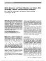
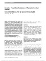
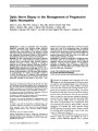
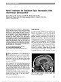
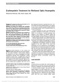
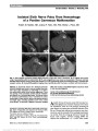
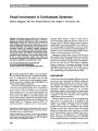
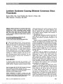
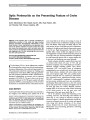
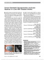
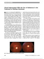
Show Vitreous Hemorrhage Secondary to Optociliary Shunt Vessels From Papilledema Clare L. Fraser, MBBS, MMed, Maysa A. Ridha, MD, Valérie Biousse, MD, Nancy J. Newman, MD Abstract: A 15-year-old adolescent girl with idiopathic intracranial hypertension was noted to have papilledema and optociliary shunt vessels. Medical management was controlling her symptoms, but vision deteriorated rapidly in the left eye secondary to a vitreous hemorrhage. Given the lack of any other cause for vitreous hemorrhage, it most likely originated from the shunt vessels. Optic nerve sheath fenestration was performed in an effort to promote regression of the papilledema and the shunt vessels. Our case illustrates a rare complication of optociliary shunt vessels in the setting of papilledema. Journal of Neuro-Ophthalmology 2012;32:332-334 doi: 10.1097/WNO.0b013e31825ba161 © 2012 by North American Neuro-Ophthalmology Society CASE REPORT Ahealthy 15-year-old adolescent girl, with normal visual function, was noted to have bilateral optic disc edema during a routine eye examination. One month later, she developed mild sporadic headaches. Magnetic resonance imaging (MRI) of the brain revealed an empty sella, flatten-ing of the posterior globes, and chronic pansinusitis. She was treated with antibiotics, and her headaches resolved, but her optic disc edema persisted. During neuro-ophthalmic evaluation 6 months later, the patient denied headaches, tinnitus, transient visual obscurations, or diplopia. She was not taking vitamin A or tetracycline derivatives. She had a body mass index of 20.1 kg/m2 without a history of recent weight gain. Her blood pressure was 100/60 mm Hg. Visual acuity was 20/25 in each eye. Color vision, slit-lamp examination, pupils, and ocular motility were normal. She had bilateral disc edema with optociliary shunt vessels (Fig. 1). Disc drusen were not detected on B-scan ultrasonography. Lumbar puncture demonstrated cerebrospinal fluid (CSF) opening pressure (OP) of 25 cm$H2O, but CSF analysis was not performed. Three weeks after lumbar punc-ture, the patient developed daily headaches requiring ibu-profen, but her examination was unchanged. One month later, she noted acute vision loss in the left eye. Visual acuity was 20/60 in the left eye with a vitreous hemorrhage FIG. 1. Bilateral papilledema with bilateral optociliary shunt vessels, more prominent on the left optic disc. Departments of Ophthalmology (CLF, MAR, VB, NJN), Neurology (VB, NJN), and Neurological Surgery (NJN), Emory University School of Medicine, Atlanta, Georgia. Supported in part by an unrestricted departmental grant (Department of Ophthalmology) from Research to Prevent Blind-ness, Inc., New York, and by National Institutes of Health/National Eye Institute core grant P30-EY06360 (Department of Ophthalmol-ogy). Dr. Newman is a recipient of the Research to Prevent Blindness Lew R. Wasserman Merit Award. Dr Clare Fraser received the Sydney Eye Hospital Alumni Travelling Fellowship 2010 and the RANZCO Eye Foundation Scholarship 2011. The authors report no conflicts of interest. Address correspondence to Nancy J. Newman, MD, Department of Neuro-Ophthalmology, Emory University, 1365 Clifton Road NE, Atlanta, GA 30322; E-mail: ophtnjn@emory.edu 332 Fraser et al: J Neuro-Ophthalmol 2012; 32: 332-334 Photo Essay Section Editor: Timothy J. McCulley, MD Copyright © North American Neuro-Ophthalmology Society. Unauthorized reproduction of this article is prohibited. (Fig. 2). Repeat lumbar puncture showed CSF OP of 28 cm$H2O and normal CSF constituents. MRI of the brain and orbits and magnetic resonance venography showed bilateral flattening of the posterior sclera, empty sella, a hypoplastic left transverse sinus, and distal right transverse sinus stenosis. The patient was begun on oral acetazolamide 250 mg twice daily. At follow-up 10 days later, she complained of postural headaches and muscle cramps. Visual acuity was 20/25, right eye, and 20/40, left eye, with bilateral optic disc edema, shunt vessels, and vitreous hemorrhage in the left eye. A left optic nerve sheath fenestration was performed 10 months after the disc edema was first noted and 1 month after her vitreous hemorrhage. One week postoperatively, visual acuity was 20/25, right eye, and 20/30, left eye, with improvement of left optic disc edema and shunt vessels. Six weeks postoperatively, visual acuities were 20/20, right eye, and 20/25, left eye, with near-complete resolution of the vitreous hemorrhage. There was slight improvement of the disc edema with persistent shunt vessels in the right eye and substantial improvement of the disc edema with decreased prominence of the shunt vessels in the left eye (Fig. 3). DISCUSSION Optociliary shunt vessels may be congenital but are more frequently reported in association with ophthalmic con-ditions that produce impaired venous outflow, including central retinal vein occlusion (1), optic nerve sheath menin-gioma (2), optic nerve glioma, optic disc drusen (3), and chronic papilledema (4). Pre-existing anastamotic capillary collaterals between the retinal and choroidal circulations undergo compensatory dilation when central retinal venous pressure is elevated (4). Flow within these shunts has been documented on the optic disc during the venous phase of fluorescein angiography and from the disc margin to vortex veins using indocyanine green angiography (5). Although vitreous hemorrhage with abnormal vessels on the optic disc is common with disc neovascularization, particularly in diabetic patients, to our knowledge, vitreous hemorrhage secondary to optociliary shunts has not been described. The combination of papilledema, disc swelling from raised intracranial pressure, and vitreous hemorrhage has been reported with Terson syndrome due to subarachnoid hemor-rhage, cerebral venous sinus thrombosis, and leukemic infiltration of the optic nerve (6). Subretinal hemorrhages in the setting of chronic papilledema from idiopathic intracranial hypertension also has been reported (7). These cases include patients found to have underlying peripapillary choroidal neo-vascular membranes (8), but none, to our knowledge, have had subretinal or vitreous hemorrhage from optociliary shunt ves-sels. Additionally, although choroidal neovascularization, disc hemorrhage, and shunt vessels have been reported in patients with disc drusen (3), we could find no published cases of vitreous hemorrhage in that setting. In our patient, vitreous hemorrhage could have occurred secondary to an unrelated condition, such as a posterior vitreous detachment, trauma, or Valsalva retinopathy, but we found no evidence of this based on history or examination. FIG. 2. Vitreous hemorrhage in the left eye. FIG. 3. Left optic disc before (A) and after (B) left optic nerve sheath fenestration. After surgery, there is reduction in optic disc edema and some regression of the optociliary shunt vessels. Vitreous hemorrhage is clearing with conservative man-agement. Fraser et al: J Neuro-Ophthalmol 2012; 32: 332-334 333 Photo Essay Copyright © North American Neuro-Ophthalmology Society. Unauthorized reproduction of this article is prohibited. Case reports have demonstrated shunt vessel regression after treatments that reduce papilledema (9-13). There are documented cases of optociliary shunt vessels disappearing in children after CSF shunting procedures (11), and in adults when intracranial pressure is reduced medically com-bined with lumbar puncture (12), or after surgical removal of a brain tumor (13). Reduction in the caliber of the optociliary shunt vessels has been shown to occur within 3 days of normalization of central retinal venous pressure (9,10). The optociliary vessels in the unoperated eye of our patient did not regress, indicating that medical management alone was inadequate to control this complication. Given that optociliary shunts from raised intracranial pressure causing papilledema may be reversible, surgical intervention with optic nerve sheath fenestration to reduce the risk of further vitreous hemorrhage may be warranted. Because vitreous hemorrhage frequently will resolve spontaneously and further hemorrhage may not occur, the risk of perma-nent vision loss from optic nerve sheath fenestration must be balanced against the risk of vision impairment either from vitreous hemorrhage or from ongoing papilledema. REFERENCES 1. Masuyama Y, Kodama Y, Matsuura Y, Sawada A, Harada K, Tsuchiya T. Clinical studies on the occurrence and the pathogenesis of optociliary veins. J Clin Neuroophthalmol. 1990;10:1-8. 2. Hollenhorst RW Jr, Hollenhorst RW Sr, MacCarty CS. Visual prognosis of optic nerve sheath meningiomas producing shunt vessels on the optic disk: the Hoyt-Spencer syndrome. Trans Am Ophthalmol Soc. 1977;75:141-163. 3. Brodrick JD. Drusen of the disc and retinal haemorrhages. Br J Ophthalmol. 1973;57:299-306. 4. Eggers HM, Sanders MD. Acquired optociliary shunt vessels in papilloedema. Br J Ophthalmol. 1980;64:267-271. 5. Muci-Mendoza R, Arevalo JF, Ramella M, Fuenmayor-Rivera D, Karam E, Cardenas PL, Recio MV. Optociliary veins in optic nerve sheath meningioma. Indocyanine green videoangiography findings. Ophthalmology. 1999;106:311-318. 6. Mayo GL, Carter JE, McKinnon SJ. Bilateral optic disk edema and blindness as initial presentation of acute lymphocytic leukemia. Am J Ophthalmol. 2002;134:141-142. 7. Orcutt JC, Page NG, Sanders MD. Factors affecting visual loss in benign intracranial hypertension. Ophthalmology. 1984;91:1303-1312. 8. Sathornsumetee B, Webb A, Hill DL, Newman NJ, Biousse V. Subretinal hemorrhage from a peripapillary choroidal neovascular membrane in papilledema caused by idiopathic intracranial hypertension. J Neuroophthalmol. 2006;26:197-199. 9. Abbasian J, Lee AG, Longmuir R, Rouleau J. Rapid regression of retinochoroidal venous collaterals following optic nerve sheath fenestration in idiopathic intracranial hypertension. Semin Ophthalmol. 2007;22:35-37. 10. Brazier DJ, Sanders MD. Disappearance of optociliary shunt vessels after optic nerve sheath decompression. Br J Ophthalmol. 1996;80:186-187. 11. Dowhan TP, Muci-Mendoza R, Aitken PA. Disappearing optociliary shunt vessels and neonatal hydrocephalus. J Clin Neuroophthalmol. 1988;8:1-8. 12. Ruth A, Newman NJ. Images in clinical medicine. Regression of optociliary shunt vessels. N Engl J Med. 2006;355:1262. 13. Tyson SL, Lessell S. Resolution of optociliary shunt vessels. J Clin Neuroophthalmol. 1986;6:205-208. 334 Fraser et al: J Neuro-Ophthalmol 2012; 32: 332-334 Photo Essay Copyright © North American Neuro-Ophthalmology Society. Unauthorized reproduction of this article is prohibited. |