| OCR Text |
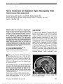
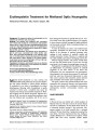
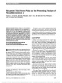
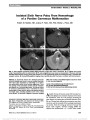
Show Rubble Rousers Matthew Rizzo, MD, FAAN In 1945 (1), Holmes found the search for anatomical substrates of visual behavior to be challenging work. The findings had to be "assembled" "with time and labor" from the "irregular rubble" of clinical material and were "rarely so simple or clean cut." I was asked to review 3 articles that recapitulate Holmes' experience: Visual involvement in corticobasal syndrome (2) and Complex visual manifestations of posterior cortical atrophy (3) that address insidious visual decline because of neuro-degenerative disease, and Bálint syndrome and visual allochiria in a patient with reversible cerebral vasoconstriction syndrome (4) that concerns sequelae of stroke. The profiles in these challenging cases may be compared to the century-old case report of Reszö Bálint (5). Bálint found a remarkable triad of visual difficulties in a man with advanced cerebrovascular disease: 1. "Spatial disorder of attention," the key component of Bálint syndrome, was described as an inability to perceive, at any one time, the several items of a visual scene. This has been compared to "visual disorientation" (6) and to "simultanagnosia," an inability to interpret the totality of a scene, despite the preserved ability to apprehend individual portions of the whole (7)-the misleading term that has stuck. Agnosia (Greek for "not knowing"), also known as "associative agnosia", is the inability to recognize previously familiar objects, despite adequate perception wherein objects are effectively stripped of their meanings. It is a memory-related disorder, a "mnestic" defect. Simulta-nagnosia is a form of "apperceptive agnosia," a failure to identify previously familiar objects as a result of impaired perception and is not memory related. (Practically speaking, patients with cerebral visual disturbances often have a mix of "mnestic" and apperceptive deficits, depending on the location and extent of lesions.) 2. "Psychic paralysis of gaze" is an inability to shift gaze voluntarily to objects of interest, despite unrestricted eye rotations. It resembles later descriptions of fixation spasm (8) and acquired ocular apraxia (apraxia, Greek for "not acting") but differs from congenital ocular motor apraxia [in which children make head thrusts occur during voluntary refixation, despite a full range of reflexive saccades (9)]. 3. Optic ataxia (OA) is the difficulty with the act of reaching under visual guidance, despite adequate limb strength and position sense. When Bálint positioned the patient's left (more impaired) hand, the man could imitate the position with his right. Note that the terms "ocular apraxia" (from the Greek, apraxia, for "not acting") and "optic ataxia" (Greek, ataktos, for "disorderly") also are ineffective in conveying the underlying mechanisms in cases such as those of Bálint. The term Bálint syndrome was coined approximately 50 years after the publication of Bálint's original report (10). Key components of the syndrome can be dissociated and also show an important overlap with the left hemineglect syndrome (11). Importantly, Bálint's patient showed prominent signs of left hemineglect syndrome with "constriction" of the "attentive field" and was unaware when approached from the left because his focus of attention was shifted 40° into the right hemifield. Postmortem examination showed a deformed, atrophic brain with bilateral lesions of the posterior parietal, upper temporal, and occipital lobes, marked damage in posterior superior and inferior parietal lobules, and changes around the left central gyrus and left internal capsule. Bálint emphasized the angular gyrus lesions, but there was damage in other vision-related structures including the posterior callosum, bilateral white matter, upper thalami, and pulvinar. The frontal lobes, and the afferent visual pathways, including optic nerve and tract, lateral geniculate body, and calcarine cortex, were largely spared. While Bálint's patient had cerebrovascular lesions, different etiologies for the syndrome complex Department of Neurology, University of Iowa, Iowa City, Iowa. Address correspondence to Matthew Rizzo, MD, FAAN, Department of Neurology, University of Iowa, 200 Hawkins Drive, Iowa City, IA 52242; E-mail: matthew-rizzo@uiowa.edu Rizzo: J Neuro-Ophthalmol 2012; 32: 299-301 299 Editorial Copyright © North American Neuro-Ophthalmology Society. Unauthorized reproduction of this article is prohibited. include neurodegenerative conditions (especially Alzheimer disease), tumor, trauma, spongiform encephalopathy, and viral infections, such as HIV, that produce differing profiles and trajectories of impairment. The report of Rajagopal et al (2) on visual involvement in presumed cortical basal syndrome (also known as cortical basal [ganglionic] degeneration [CBD]) in a 60-year-old woman with progressive vision and limb control difficulties had to be distinguished from Bálint syndrome. The CBD symptom complex includes "alien limb" (or hand), limb apraxia, cortical sensory loss, focal reflex myoclonus, rigid-ity, akinesia, postural action tremor, limb dystonia, hyper-reflexia, and postural instability (12). Unlike OA, the alien limb phenomenon is not limited to reaching under visual guidance (11). The hand seems to move as if possessed by an outside agent and may show a grasp reflex and ele-ments of "magnetic apraxia" (13) in association with frontal lobe lesions. Whereas OA may occur as part of Bálint syn-drome in Alzheimer disease (with amyloid and tau pathol-ogy) (14), alien hand syndrome is more characteristic of CBD, a tauopathy (15). The patient of Rajagopal et al (2) experienced frequent involuntary movements of the upper body and occasional tonic flexed posturing of the left arm (and had no resting tremor to suggest Parkinson disease). Yet, we cannot be sure of the underlying behavioral impairments of disease in this patient. She showed "marked ideomotor apraxia" (this gen-erally manifests as an inability to imitate hand gestures or to pantomime use of tools [e.g., comb, toothbrush]); yet, it is not clear how this was defined or tested. She tended to use her left hand much less than the right, as can occur with motor neglect-which was apparently not considered. Could she imitate the position of one hand passively posi-tioned by the examiner, with the other hand (see Bálint, above)? This would help exclude a role of cortical sensory loss or dorsal column disease in disordered limb control. The patient also seemed to show "profound simultagno-sia" based on her poor description of the "Cookie Thief [sic]" picture. (The Cookie Theft picture originated from the Boston Diagnostic Aphasia Examination (16) not from the National Institutes of Health as Rajagopal et al (2) and Walsh et al (4) indicate). Yet, "her cognitive impairment prevented prolonged coherent speech," so her inability to describe the Cookie Theft could have been language related. The patient denied loss of vision, photopsia, scotoma, or diplopia but had severe communication problems and dementia. Yet, denial of defects because of the lack of self-awareness of impairment (known as "anosognosia") is common in dementia. An earlier Mini Mental Status (dementia screening) score was 12/30 (very impaired), and dementia could have progressed even further by the time the authors saw the patient; yet, this was apparently not tested. She could not cooperate for visual field assessment, which means we cannot exclude visual field defects causing what might look like, it but is not simultanagnosia (e.g., objects "disappearing" into undetected scotomata). The authors do not state what medications the patient was taking or how they excluded posterior cortical atrophy (PCA), multisys-tems atrophy, B12 deficiency, syphilis, vasculitis, and other etiologies. Magnetic resonance imaging (MRI) showed "dif-fuse cortical atrophy, with some predominance in the pos-terior parietal regions," which fits PCA (better than CBD), but we do not get to see the MRI for ourselves. Abnormal signal in basal ganglia could occur with metal deposition (copper in Wilson disease, iron in Hallervorden-Spatz), although these diagnoses seem unlikely. Functional brain imaging may have helped disentangle this case (see Rene et al (3)). Position emission tomography (PET) or single-photon emission computed tomography (SPECT) might show asymmetric activation in subcortical (basal ganglia) and cortical (frontal-parietal) regions in CBD (15). Rene et al (3) report 5 cases of PCA, a visual variant of Alzheimer disease (17), in which visual difficulties are not explained by ocular pathology. Three patients had homon-ymous hemianopia, and 3 showed right posterior atrophy on MRI. Diagnosis was aided by observing clinical signs and PET in 2 patients with nonspecific MRI findings. Aspects of 1 case resembled the cases of both Rajagopal et al (parkinsonism or basal ganglionic features) and Walsh et al (right-left confusion). This patient had left homony-mous hemianopia, left upper limb neglect and deafferenta-tion, alexia, agraphia, right-left confusion, finger agnosia, bilateral stereoagnosia, sensory extinction, OA, and mild par-kinsonism. SPECT showed severe right parietotemporo-occipital hypoperfusion. Walsh et al (4) reported "visual allochiria" in a 67-year-old woman with Bálint syndrome. MRI initially showed acute right parietal-occipital infarction and bilateral parietal-occipital infarctions 9 days later. Impaired left arm move-ment under visual guidance was not explained to the degree of weakness. Allochiria (Greek for "other hand") has been observed following right parietal lobe lesions (18). Patients mislocalize the side of the body that has been stimulated, despite adequate sensation. Walsh et al report allochiria in vision, but it is unclear how they measured this defect or distinguished it from (apperceptive) the effects of simultanag-nosia (e.g., from irregular shifts of attention between hemi-fields), extinction to double simultaneous stimulation (generally on the left in patients with right parietal lesions), or akinetopsia-as in a patient reported by Zihl et al (19), who had bilateral parietal lesions and complained of objects appearing on one side or another without seeing them move in between (because of her profound motion apperception). The patient of Walsh et al may have had Gerstmann syndrome as a result of her left parietal lesion, which includes right-left confusion (20). Certain drugs that may have affected motor tone and cognition, such as promethazine, hydrocodone, alprazolam, and atenolol, further confound this case. Visual and neuropsychological assessments are lacking. 300 Rizzo: J Neuro-Ophthalmol 2012; 32: 299-301 Editorial Copyright © North American Neuro-Ophthalmology Society. Unauthorized reproduction of this article is prohibited. Visual disturbances caused by brain lesions, as above, offer a unique window on the psychoanatomical substrates of human behavior. The brain constructs a seamless, detailed picture from partial glimpses of the visual world. Brain lesions in Bálint syndrome destroy this illusion, pro-ducing a piecemeal visual experience (simultanagnosia/ visual disorientation) and impaired visual control of eye and hand movements (ocular apraxia and optic ataxia). Evi-dence from these cases remains highly relevant to under-standing the neural substrates of vision, motor control, memory, and even consciousness. Because Bálint syndrome is not common and is difficult to assess with standard clinical tools, the literature is dom-inated by case reports. However, it is risky to generalize from these historical or modern single case reports, which are confounded by case selection bias, nonuniform application of operational definitions, inadequate study of basic vision, poor lesion localization, and failure to distinguish between deficits in the acute and chronic phases of recovery or dura-tion or stage of neurodegeneration. There is variability of lesion effects, and the bilateral brain lesions in these cases cause extensive visual and cognitive impairments that hinder clinical testing. Interesting abnormalities are much more likely to be reported and less striking ones ignored, creating biased profiles. Lesion effects and recovery vary with patient age, time since lesion onset, and white matter involvement. Patients studied in the acute phase tend to show more pro-found deficits than those studied months or years later in the chronic phase of recovery. Methodological concerns include failure to adequately assess basic visual functions (other than acuity) and to consider confounding eye conditions (such as retinopathy, cataracts, or optic neuropathy). In addition, the abnormalities of eye or hand movements in the above cases were not quantified in any way. Cognitive and visual testing were sparse, neuroimaging data were limited, operational definitions were unclear, and theoretical interpretations are lacking. Future clinical research studies should address pos-sible underlying psychoanatomical ("bottom up" and "top down") mechanisms, with specific consideration to visual working memory and attention (including spatial and object attention) and to systems for the identification of object structure and depth from binocular stereopsis, kinetic depth, motion parallax, eye movement signals, and other cues (11). The successful approach to these cases requires clear opera-tional definitions of behavioral impairments and sufficiently detailed assessments to classify patients who, as Holmes recognized, may be challenging to test because of their visual and cognitive difficulties. REFERENCES 1. Holmes G. Ferrier lecture. The organisation of the visual cortex in man. Proc R Soc Ser B. 1945;132:348-361. 2. Rajagopal R, Bateman R, Van Stavern GP. Visual involvement in corticobasal syndrome. J Neuroophthalmol. 2012;32:xx-xx. 3. Reñá R, Muñoz S, Campdelacreu J, Gascon-Bayarri J, Rico I, Juncadella M, Arrunga J. Complex visual manifestations of posterior cortical atrophy. J Neuroophthalmol. 2012;32:xx-xx. 4. Walsh RD, Floyd JP, Eidelman BH, Barrett KM. Bálint syndrome and visual allochiria in a patient with reversible cerebral vasoconstriction syndrome. J. Neuroophthalmol. 2011 Nov 13 [epub ahead of print]. 5. Bálint R. Seelenlähmung des "Schauens", optische Ataxie, räumliche Störung der Aufmerksamkeit. Monatschrift für Psychiatrie und Neurologie. 1909;25:51-181. 6. Holmes G. Disturbances of visual orientation. Br J Ophthalmol. 1918;2:449-468. 7. Wolpert I. Die Simultanagnosie. Zeitschrift fur die gesamte. Neurologie und Psychiatry. 1924;93:397-415. 8. Johnston JL, Sharpe JA, Morrow MJ. Spasm of fixation: a quantitative study. J Neurol Sci. 1992;107:166-171. 9. Cogan DG. Congenital ocular motor apraxia. Can J Ophthalmol. 1965;1:253-260. 10. Hécaen H, de Ajuriaguerra J. Bálint's syndrome (psychic paralysis of visual fixation) and its minor forms. Brain. 1954;77:373-400. 11. Rizzo M, Vecera SP. Psychoanatomical substrates of Bálint's syndrome. J Neurol Neurosurg Psychiatry. 2002;72:162-178. 12. Riley DE, Lang AE, Lewis A, Resch L, Ashby P, Hornykiewicz O, Black S. Cortical-basal ganglionic degeneration. Neurology. 1990;40:1203-1212. 13. Denney-Brown D, Chambers RA. Physiological aspects of visual perception. I. Functional aspects of visual cortex. Arch Neurol. 1976;33:219-227. 14. Wray S, Noble W. Linking amyloid and tau pathology in Alzheimer disease: the role of membrane cholesterol in Ab-mediated tau toxicity. J Neurosci. 2009;29:9665-9667. 15. Rizzo G, Martinelli P, Manners D, Scaglione C, Tonon C, Cortelli P, Malucelli E, Capellari S, Testa C, Parchi P, Montagna P, Barbiroli B, Lodi R. Diffusion-weighted brain imaging study of patients with clinical diagnosis of corticobasal degeneration, progressive supranuclear palsy and Parkinson's disease. Brain. 2008;131:2690-2700. 16. Goodglass H, Kaplan E. The Assessment of Aphasia and Related Disorders, 2nd edition. Philadelphia, PA: Lea & Febiger, 1983. 17. Benson DF, Davis RJ, Snyder BD. Posterior cortical atrophy. Arch Neurol. 1988;45:789-793. 18. Meador KJ, Allen ME, Adams RJ, Loring DW. Allochiria vs allesthesia-is there a misperception. Arch Neurol. 1991;48:546-549. 19. Zihl J, von Cramon D, Mai N. Selective disturbance of movement vision after bilateral brain damage. Brain. 1983;106:313-340. 20. Vallar G. Spatial neglect, Balint-Holmes' and Gerstmann's syndrome, and other spatial disorders. CNS Spectr. 2007;12:527-536. Rizzo: J Neuro-Ophthalmol 2012; 32: 299-301 301 Editorial Copyright © North American Neuro-Ophthalmology Society. Unauthorized reproduction of this article is prohibited. |