| OCR Text |
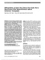
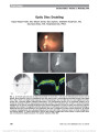
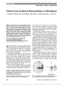
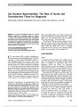
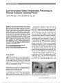
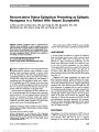
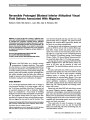
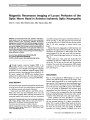
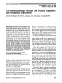
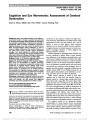
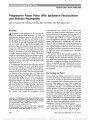
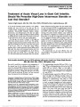
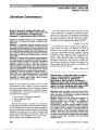
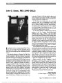
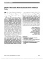
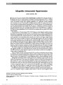
Show Primary Dural Lymphoma Masquerading as a Meningioma Kaushal M. Kulkarni, MD, Linda Sternau, MD, Sander R. Dubovy, MD, Byron L. Lam, MD Abstract: A 39-year-old woman noted progressive blurred vision in the right eye for 1 year. The right eye had visual acuity of 20/25, an afferent pupillary defect, pale optic nerve, and cecocentral scotoma. Magnetic resonance imaging findings were consistent with en plaque meningi-oma of the planum sphenoidale, which encircled the right optic nerve at the optic canal. The tumor was internally debulked to preserve the optic nerve. Histopathologic and molecular analysis revealed a low-grade B-cell lymphoma. Further evaluation showed no evidence of systemic dis-ease. Primary dural lymphomas are a distinct entity that may mimic meningioma and cause vision loss. Journal of Neuro-Ophthalmology 2012;32:240-242 doi: 10.1097/WNO.0b013e31825103a5 © 2012 by North American Neuro-Ophthalmology Society A39-year-old woman noted painless, progressive blurred vision in the temporal aspect of her right visual field for 1 year. Medical history was significant for hypertension treated with enalapril. The woman was a professional banker, entertainer, and singer from Trinidad. She denied the use of tobacco or illicit drugs. Visual acuity was 20/25, right eye, and 20/20, left eye. She had diminished color vision in the right eye with a 0.6 log unit relative afferent pupillary defect. Extraocular movements were full, and external and anterior segment examinations were unremarkable. Intraocular pressure was 19 mm Hg bilaterally. Automated visual field of the right eye showed central and temporal loss (Fig. 1) while the left field was normal. There was mild optic disc pallor in the right eye, and the left fundus was normal. The results of brain magnetic resonance imaging (MRI) were felt to most likely represent en plaque meningioma of the planum sphenoidale, compressing the right optic nerve with involvement of the right optic canal, orbital apex, and cavernous sinus with extension to the left optic canal (Fig. 2A-C). Other diagnostic considerations included a lymphoproliferative disorder, sarcoidosis, and nonspecific inflammation (pseudotumor). Three days later, a right fronto-orbital craniotomy was performed to debulk the tumor. The surgical findings appeared typical of an en plaque meningioma (Fig. 3). The pathologic specimen revealed an infiltrate of small basophilic cells (Fig. 4A). Staining for epithelial membrane antigen was negative for cells of meningoepithelial origin. CD20 stained the majority of the cells, indicating a B-cell proliferation with a few interspersed reactive T cells (Fig. 4B). The cells were highlighted by CD138, indicating plasmacytoid differentiation (Fig. 4C). Ki67, a nuclear protein only present during active phases of the cell cycle, showed a moderate proliferative index (Fig. 4D). Molecu-lar analysis and polymerase chain reaction detected immu-noglobulin (IGH and IGk) gene rearrangements. The histology and genetic results confirmed the diagnosis of B-cell lymphoma. Staging, including bone marrow biopsy and whole-body positron emission tomography-computed tomography, showed no evidence of systemic disease. The patient was treated with 45 Gy of intensity-modulated radiation ther-apy. Eighteen months later, visual acuity was 20/25, right eye, and 20/20, left eye. The right visual field deficit FIG. 1. Automated visual field of the right eye shows a cecocentral scotoma and temporal field depression. Department of Ophthalmology (KMK, SRD, BLL), Bascom Palmer Eye Institute, University of Miami, Miami, Florida; and Department of Neurosurgery (LS), Memorial Neuroscience Center, Memorial Regional Hospital, Hollywood, Florida. The authors report no conflicts of interest. Address correspondence to Kaushal M. Kulkarni, MD, 900 NW 17th Street, Miami, FL 33139; E-mail: kaushal.kulkarni@gmail.com 240 Kulkarni et al: J Neuro-Ophthalmol 2012; 32: 240-242 Photo Essay Section Editor: Timothy J. McCulley, MD Copyright © North American Neuro-Ophthalmology Society. Unauthorized reproduction of this article is prohibited. remained stable. The MRI showed successful debulking of the tumor with no evidence of recurrence (Fig. 2D-F). Lymphomas arising primarily from the meninges without brain or systemic involvement are rare and differ biologically from other central nervous system (CNS) lymphomas (1,2). When they do occur, they typically consist of a low-grade B-cell lymphoma (1), as in our case. Primary dural lymphoma (PDL) typically has an indolent course and generally can be cured with local therapy (3). It differs from other types of CNS lymphoma (either primary or metastatic), which are typically aggressive, high-grade neoplasms with a poor prognosis. The pathogenesis of PDL is not well understood. The dura is devoid of lymphoid tissue, as opposed to the gastrointestinal tract, where histologically similar marginal zone lymphomas typically originate from chronic antigenic stimulation (1). It has been hypothesized that meningoepithelial cells, which are present throughout the arachnoid membrane, may give rise to these neoplasms in a similar fashion (2). PDL is initially often diagnosed as meningioma because of its virtually identical MRI features, including a dural tail (4). Thus, in cases of suspected meningioma, it is prudent to obtain a pathologic specimen whenever possible or clinically warranted. FIG. 2. Contrast-enhanced T1 axial (A) and coronal (B, C) magnetic resonance imaging (MRI) with fat suppression. A. Tumor involves the orbital and canalicular portions of the right optic nerve and the anterior clinoid process. B. There is enhancement of the right orbital apex (arrow) and meninges of the floor of the anterior cranial fossa (arrowheads). C. Tumor enhancement is seen below the prechiasmal optic nerves (arrows) with encasement of the right intracavernous internal carotid artery. D-F. Eighteen months after treatment, MRI shows successful resolution of the tumor with no evidence of recurrence. FIG. 3. Intraoperative photographs before (A) and after (B) surgical debulking. *Olfactory nerve; arrow, area where anterior clinoid and tumor were removed; arrowhead, falciform ligament; O, optic nerve; S, Surgicel (Ethicon, Somerville, NJ), where tumor over the tuberculum sellae was removed; T, tumor. Kulkarni et al: J Neuro-Ophthalmol 2012; 32: 240-242 241 Photo Essay Copyright © North American Neuro-Ophthalmology Society. Unauthorized reproduction of this article is prohibited. If a lymphomatous neoplasm is suspected on the initial pathol-ogy, further testing should be done to identify the precise histologic subtype. A meticulous search for an occult source is critical because this will determine treatment and prognosis. REFERENCES 1. Iwamoto FM, Abrey LE. Primary dural lymphomas: a review. Neurosurg Focus. 2006;21:E5. 2. Kumar S, Kumar D, Kaldjian EP, Bauserman S, Raffeld M, Jaffe ES. Primary low-grade B-cell lymphoma of the dura: a mucosa associated lymphoid tissue-type lymphoma. Am J Surg Pathol. 1997;21:81-87. 3. Puri DR, Tereffe W, Yahalom J. Low-dose and limited-volume radiotherapy alone for primary dural marginal zone lymphoma: treatment approach and review of published data. Int J Radiat Oncol Biol Phys. 2008;71:1425-1435. 4. Sotoudeh H, Yazdi HR. A review on dural tail sign. World J Radiol. 2010;2:188-192. FIG. 4. Histopathologic specimen. A. The tumor is composed primarily of small basophilic cells (hematoxylin and eosin, ·200). B. Immunostaining shows a lymphoid cell population with predominance of CD20+ B cells (brown) and few interspersed CD3+ T cells (red) (·400). C. Many cells stain positive for CD138, indicating plasmacytoid differentiation (·400). D. Staining for Ki67 shows a moderate proliferative index (·200). 242 Kulkarni et al: J Neuro-Ophthalmol 2012; 32: 240-242 Photo Essay Copyright © North American Neuro-Ophthalmology Society. Unauthorized reproduction of this article is prohibited. |