| OCR Text |
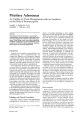
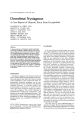
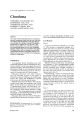
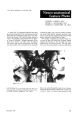
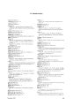
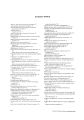
Show ]. Clin. Neuro-ophthalmol. 3: 251-257,1983 Chordoma TOSHIHIRO TAKAHASHI, M.D. TOSHIMICHI ASAI, M.D. YOSHIMASA ISAYAMA, M.D. NORIHIKO TAMAKI, M.D. SATOSHI MATSUMOTO, M.D. Abstract Four cases of intracranial chordomas are reported. Intracranial chordomas initially tend to occur with sixth nerve paresis and subsequently with visual disturbance and optic disc changes. Computed tomography demonstrated an isodense mass in one case, a lowdensity mass in another case, and both an isodense and a partial high-density in two cases. Histopathologically, isodense areas were coincident with regions of compact tumor cells, low-density areas coincided with abundant mucinous intercellular matrices, and high-density areas were coincident with cartilaginous matrices or calcifications. Introduction As is generally known, chordomas arise from notochordal remnants and are classified into intracranial, vertebral, and sacrococcygeal sites according to their location. The world literature, reviewed by Utene and Pughl in 1955, mentioned 550 cases of chordomas in the following locations: 197 intracranial, 81 vertebral, and 227 sacrococcygeal. Intracranial chordomas occur rarely; Schisano and Tovi2 described 10 cases of this tumor in Olivecrona's series of 6,700 cases of verified brain tumors, and Trappe and Weidenbach3 reported only one case in 3,440 intracranial tumors. Although intracranial chordomas often initially manifest ocular signs and symptoms, only several papers on this disease have appeared in the field of ophthalmology. Bagan and Hollenhorst4 analyzed ocular manifestations in 63 cases of intracranial chordomas. Other reports described a single case each.3 . 5-8 The relationship between computed tomographic and histopathological findings of this tumor still remains to be elucidated. This paper deals with four cases of histopathologically proven intracranial chordomas and ana- From the Department of Ophthalmology (TT, TA, YI) and the Department of Neurosurgery (NT, SM), School of Medicine. Kobe University, Kobe, Japan. December 1983 lyzes the computed tomographic findings in relation to the histopathological aspects of this tumor. Case Reports Case I A 39-year-old male was referred to us on July 27, 1975, because of intermittent diplopia for 1 month. The vision was 20/16 in each eye. A mild right sixth nerve paresis was observed. Otherwise, anterior segments, media, and ocular fundi were normal. On follow-up study on September 22, 1975, a mild left sixth nerve paresis was also found. CT scans showed a prepontine mass, consisting of an isodensity in relation to the normal brain and a partial high-density. An isodense area enhanced inhomogenously on the contrast study (Fig. 1). Subtotal resection of the tumor was performed on November 27, 1975. Histopathological studies confirmed a chordoma. The tumor cells with vacuolated cytoplasms were arranged compactly in the mucinous intercellular matrix. In some places the tumor cells were present in the hyaline cartilaginous matrix (Fig. 2). Postoperatively, a course of conventional radiation therapy (5,000 rads) was carried out, and the patient made a partial recovery with minimal bilateral sixth nerve paresis for 4 years. Thereafter, left fifth and sixth nerve palsies, as well as nausea, vomiting, and papilledema due to increased intracranial pressure, occurred. The patient died on October 2, 1980. Case 2 A 51-year-old male was seen on January 25, 1978, because of diplopia and headache since 1974 and progressive visual disturbance for 3 months. The corrected vision was 20/20 in the right eye and 20/40 in the left eye. A mild right sixth nerve paresis was detected. Otherwise, anterior segments and media were normal. Ophthalmoscopy revealed a normal optic disc in the right eye and slight simple optic atrophy in the left eye. Goldmann perimetry demonstrated bitemporal hemianopia (Fig. 3). CT scan showed a round, suprasellar iso- 251 Chordoma Figure 1. Case I-left: Prepontine mass consisting of an isodensity .:md partial high-density on CT scan. Right: Inhomogenous enhancement on contrast study. '. Figure 2. Case I-Tumor cells in the hyaline cartilaginous matrix. (H&E X100.) 252 Journal of Clinical Neuro-ophthalmology Takahashi et al. -7 ===-~_~ // .-------- Figure 3. Case 2-Bitemporal hemianopia on Goldmann visual fields. Figure 4. Case 2-1eft: A suprasellar, isodense mass on CT scan. Right: Marked enhancement on contrast study. dense mass. The lesion enhanced markedly on the contrast study (Fig. 4). Subtotal resection of the tumor was carried out on March 13, 1978, and the tissue examination confirmed a chordoma. The neoplastic cells were arranged in lobulates or alveolar structures in the matrix and stained positively with mucicarmine (Fig. 5). Many vessels were found in the lesion. Subsequent irradiation of 4,000 rads was given. The right sixth nerve paresis disappeared and the visual function remained stable. Case 3 A 31-year-old female was seen on August 22, 1979, because of progressive diplopia for 3 years. The corrected vision was 20/16 in each eye. A complete left sixth nerve palsy was observed. Otherwise, anterior segments, media, and ocular December 1983 fundi were normal. CT scans showed a large, lowdensity mass in the left middle fossa. The margin of the lesion enhanced by intravenous contrast (Fig. 6). Subtotal resection of the tumor was performed on October 8, 1979. Histopathological studies confirmed a chordoma. The tumor cells with intracytoplasmic vacuoles were arranged in cords in the abundant amorphous matrix (Fig. 7). The tumor was pseudoencapsulated by the fibrovascular tissues. Postoperatively, a course of Cobalt'ill irradiation (6,000 rads) was given. The left sixth nerve palsy persisted. Case 4 A 21-year-old female was seen on December 17, 1980, because of diplopia for 3 months. The cor- 253 Chordoma . .. Figure 5. Case 2-Tumor cells of alveolar structures in the matrix stained with mucicarmine. (Mucicarmine stain, XlOO.) Figure 6. Case 3-left: low-density mass in the left middle fossa on cr scan. Right: Enhancement in the margin of the lesion on contrast study. rected vision was 20/20 in each eye. Partial bilateral sixth nerve pareses were detected. Otherwise, ocular examination revealed no remarkable changes. CT scans showed a prepontine mass having both isodense and high-density components. The lesion was inhomogenous and enhanced markedly on the contrast study (Fig. 8). 254 Partial resection of the tumor was carried out on February 26, 1981. The tissue examination demonstrated a chordoma. Tumor cells were present in the hyaline matrix, occasionally accompanied by granular calcifications (Fig. 9). Postoperatively, irradiation of 4,400 rads was given. Bilateral sixth nerve pareses were improved Journal of Clinical Neuro-ophthalmology Takahashi et al. .. Figure 7. Case 3-Physaliphorous tumor cells in the abundant amorphous matrix. (H&E XIOO.) Figure 8. Case 4-left: Prepontine mass having an isodensity and a partial high-density on CT scan. Right: Inhomogenous, marked enhancement on contrast study. temporarily. Follow-up study on February 18, 1983, revealed complete bilateral sixth nerve palsies and moderate right third and fourth nerve paresis. The corrected vision was 20/200 in the right eye and 20/20 in the left eye. Ophthalmoscopy revealed slight simple optic atrophy in the right eye and a normal optic disc in the left eye. Irradiation of 3,600 rads was given. December 1983 Discussion Intracranial chordomas can occur at any age and may particularly manifest symptoms during the third and fourth decades of life. Male involvement was predomm. ant .4.9-11 In our study, the ages ranged from 21 to 51 years, averaging 35.5 years; and the male to female ratio was 1:1. 255 Chordoma '. Figure 9. Case 4-Tumor cells in the hyaline matrix with granular calcifications. (H&E X100.) Symptoms of intracranial chordomas reported in the order of decreasing frequency were headache, diplopia, decreased visual acuity, visual field defect, ataxia, dysphagia, decreased hearing, endocrine disturbance, facial numbness, weakness of facial muscles, nausea, vomiting, and papilledema. 4 . 1O-1~ In the present study, unilateral or bilateral sixth paresis was observed initially in all four cases; thereafter, visual disturbance in two cases, simple optic atrophy in two cases, papilledema in one case, and third, fourth, and fifth nerve pareses in one case. Therefore, intracranial chordomas initially tended to occur with sixth nerve paresis and subsequently with visual disturbances and/or optic disc changes. Several papers on CT scans concerning intracranial chordomas have appeared in the literature" ~ and have reported various findings on the nonenhanced and contrast studies. In the present study, CT scans demonstrated a mass having an isodensity and a partial high-density in two cases, an isodense mass in one case, and a low-dense mass in one case. The lesions enhanced to different degrees on the contrast studies. A correlation between CT and histopathological findings was as follows: isodense areas were coincident with regions of compact tumor cells, low-density areas coincided with abundant mucinous intercellular matrices, and high-density areas were coincident with cartilaginous matrices or calcifications. Fur-thermore, the various degrees of enhancement on the contrast studies depended upon the vascularity in the tumors and pseudocapsules. References 1. Utene, J.R., and Pugh, D.G.: The roentgenologic aspects of chordomas. Am. J. RoentgenoJ. 74: 593608,1955. 2. Schisano, G., and Tovi, D.: Clivus chordomas. Neurochirurgica 5: 99-120, 1962. 3. Trappe, A., and Weidenbach, W.: Einseitiger Exophthalmus und temporare gleichseitige Okulomotoriusparese als einzige Symptome eines ausgedehnten Chordoms der Schadelbasis. KJin. Monatsbl. Augenheilkd. 171: 953-958,1977. 4. Bagan, S.M., and Hollenhorst, R.W.: Ocular manifestation of intracranial chordomas. Trans. Am. OphthaJmol. Soc. 78: 148-155, 1980. 5. Daiker, B.C.: Chordom der Orbita. Ophthalmologica 176: 236-239, 1978. b. Ferry, A.P., Haddad, H.M., and Goldmann, J.L.: Orbital invasion by an intracranial chordoma. Am. ]. OphthalmoJ. 92: 7-12, 1981. 7. Kline, l.B., and Glaser, J.5.: Bilateral abducens nerve palsies from clivus chordoma. Ann. OphthaJmol. 13: 705-707,1981. 8. Neetens, A., Bultinck, R., Martin, J.L and Solheid, J.: Intrasellar adenoma and chordoma. Neuro-ophthaJmology 2: 123-135, 1980. 9. Dahlin, D.C., and Mac Carty, C.S.: Chordoma, a study of fifty nine cases. Cancer 5: 1170-1178, 1952. 256 Journal of Clinical Neuro-ophthalmology 10. Heffelfinger, M.j., Dahlin, D.C, Mac Carty, CS., and Beabout, J,W.: Chordomas and cartilaginous tumors at the skull base. Cancer 32: 410-420,1973. 11. Karmin, R.P., Potanos, J,N., and Pool, j.L.: An evaluation of the diagnosis and treatment of chordoma. ]. Neurol. Neurosurg. Psychiatry 27: 157-165, 1964. 12. Falconer, M.A., Bailey, I.C, and Duchen, L.W.: Sur- December 1983 Takahashi et al. gical treatment of chordoma and chondroma of the skull base. ]. Neurosurg. 29: 261-274, 1968. Write for reprints to: Toshihiro Takahashi, MD., Department of Ophthalmology, School of Medicine, Kobe University, Kusunoki-cho, 7-chome, Chuo-ku, Kobe (650), japan. 257 |