| OCR Text |
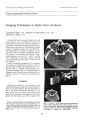
Show Journal of Clinical Neuro-ophtlmlmology 8(4): 273-276, 1988. Literature Abstracts To Image Or Not To Image. Gittinger JW Jr. With comments by JL Keltner, NR Miller, and RM Burde. Surv Ophthalmol 1988;32:35~6 (Mar-Apr). [No reprints available.] This clinical pathological conference involves an 8-year-old girl with poor vision documented since age 4 years and nystagmus. She had a mass on computerized tomographic scanning purportedly involving chiasm, hypothalamus, and cerebral peduncles (but a rather poor quality computerized tomographic scan is presented with the article). The discussants do their usual thoughtful and compulsively exhaustive job discussing this case and its management. The controversies regarding treatment of chiasmal glioma, if any can be recommended, are stressed. This is good reading to bring yourself up to date regarding the management of such lesions. LYI1 A. Sedwick, M.D. Posterior Scleritis. Benson WE. Surv Ophthalmol 1988;32:297-316 (Mar-Apr). [Reprint requests to Dr. W. Benson, Retinovitreous Associates, 910 E. Willow Grove Ave., Philadelphia, PA 19118.] This paper sets out to review the world literature on posterior scleritis and describes findings in 43 patients from Wills Eye Hospital. Similarities between this disease and orbital pseudotumor are discussed. Many nice clinical photos, ultrasound, and one computerized tomographic scan are presented as well as good summary tables comparing and contrasting posterior scleritis with other similar disorders. This may not be the most fascinating neuro-ophthalmic subject around but this is too nice a review to pass up. LYI1 A. Sedwick, M.D. Imaging of Cerebral Blood Flow and Metabolism in Amblyopia by Positron Emission Tomography. Derner JL, von Noorden GK, Volkow NO, Gould 273 {~, 1988 Raven Press, Ltd., New York KL. Am J Ophthalmol 1988;105:337-47 (April). [Reprint requests to Dr. J. L. Derner, Cullen Eye Institute, Baylor College of Medicine, 6501 Fannin St., NC-200, Houston, TX 77030.] Positron emission tomography (PET scanning) was performed in four patients with unilateral childhood amblyopia and two normals. In the normals, either eye gave similar PET evidence of stimulation of primary and accessory visual cortex, but in all amblyopic patients, the sound eye stimulated primary and accessory visual cortex more than the amblyopic eye. This is a nicely detailed study and includes colored PET images. Lyn A. Sedwick, M.D. Optic Nerve Swelling Secondary to the Obstructive Sleep Apnea Syndrome. Bucci FA, Krohel GB. Am JOphthalmoI1988;105:428-30 (April). [Inquiries to Dr. G. B. Krohel, Department of Ophthalmology, Albany Medical College, Albany, NY 12208.] A patient with obstructive sleep apnea had normal visual function but bilateral disk edema that cleared with permanent tracheostomy. Lumbar puncture showed an opening pressure of 170 mm H20 and computerized tomographic scanning demonstrated mild swelling of the right optic nerve. Increased venous pressure is the presumed cause of disk edema, and usually increased intracranial pressure is present. Lyn A. Sedwick, M.D. Botulinum Toxin A-Induced Protective Ptosis in Corneal Disease. Kirkness CM, Adams GGW, Dilly PN, Lee JP. Ophthalmology 1988;95:47~80 (April). [Reprint requests to Dr. C. M. Kirkness, Moorfields Eye Hospital, City Rd., London, England EC1V 2PD.] These authors used botulinum toxin A at the dose usually given to a single extraocular muscle in stra- |