| OCR Text |
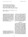
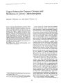
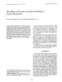
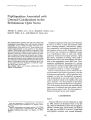
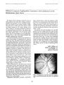
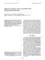
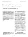
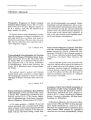
Show tournai < lr Clinical Neliro- opJrtJralmoloS. 1f 11( 3): 175- 180, 1991. Papillopathies Associated with Unusual Calcifications in the Retrolaminar Optic Nerve Alfredo A. Sadun, M. D., Ph. D., Ronald L. Green, M. D., Janis R. Nobe, M. D., and Miriam R. Cano, M. D. < 91991 Raven Press, Ltd., New York We examined three patients with optic disc edema and peripapillary hemorrhages. Each was found by standard echography to have calcified nodules within the retrobulbar portion of the optic nerves. These nodules were located approximately 2 mm posterior to the lamina cribrosa. Each patient had unilateral congestion of the optic nerve head with dilated, tortuous retinal veins that appeared much like a partial central retinal vein occlusion; one patient subsequently developed optic atrophy. The central location of the calcifications within the anterior aspect of the optic nerves suggests that each nodule may have been situated within the central retinal vein or artery. Calcifications within the retrolaminar space may be associated with some etiologies of unilateral congestion of the optic nerve head. Key Words: Optic nerve head- Ultrasound- Disc edema- Calcifications. From the Department of Ophthalmology, University of Southern California School of Medicine, Los Angeles, Cahfornia, U. S. A. Address correspondence and reprint requests to Dr. Alfredo A. Sadun at Department of Ophthalmology, USC School of Medicine, 1355 San Pablo Street, Los Angeles, CA 90033, U. S. A. 175 Unilateral congestion of the optic nerve head can be seen in a variety of local and systemic conditions, including metabolic, inflammatory, infiltrative, compressive, and ischemic processes ( 1). Ocular diseases such as uveitis, hypotony, and central retinal vein occlusion are additional causes ( 1,2). Papilledema refers to optic disc edema caused by increased intracranial pressure. Optic disc edema, regardless of etiology, often leads to dilated retinal veins with hemorrhages and infarctions of the peripapillary nerve fiber layer. Thus, congestive papillopathy presenting as a picture consistent with partial or impending central retinal vein occlusion can refer to disc edema with vascular dilatation caused by a variety of etiologies. Herein we describe three cases in which the clinical presentation and fundus appearance were that of congestive papillopathy with peripapillary hemorrhages. In each case standardized echography ( 3,4), utilizing both A- scan and contact B- scan, revealed a calcified nodule in the middle of the optic nerve, located 2 ± 0.5 mm posterior to the lamina cribrosa ( as measured from the B- scan echograms). Two of the patients had fundi that maintained the appearance of unilateral optic disc edema with dilated, tortuous retinal veins. The third patient ultimately developed severe optic atrophy. CASE REPORTS Case 1 A 24- year- old man was first examined on April 23, 1986, because of decreased vision in his left eye, which he attributed to blunt trauma against a cabinet 2 months prior. Visual acuity was 20/ 20 in the right eye and 20/ 30- 2 in the left. Visual fields and pupillary reactions were normal bilaterally. 176 A. A. SADUN ET AL. Anterior segment examination showed no abnormalities, and intraocular pressure was 18 mm Hg bilaterally. The right fundus appeared normal. Ophthalmoscopy of the left eye revealed extensive perivenous flame- shaped hemorrhages, venous dilatation, and papillary swelling. There was no discernible optic disc cup in either eye. Laboratory studies, including complete blood count, blood chemistries, and erythrocyte sedimentation rate showed normal values. Antinuclear antibody titer, rheumatoid factor, VORL, and tuberculin skin test were all nonreactive. A chest film showed no abnormalities. Three months later the visual acuity in the left eye had decreased to 20/ 200, and an afferent pupillary defect was present. Color vision testing using Ishihara color plates revealed 618 correctly identified with the right eye and 4/ 8 with the left eye; brightness sense was decreased to 50% in the left eye ( 5). Fundus examination of the left eye revealed papillary swelling with dilated disc vessels and hyperemia, tortuous and dilated retinal veins, hard exudates in the macula, and scattered hemorrhages throughout the posterior pole ( Fig. 1). Visual field testing of the left eye with a Goldmann perimeter failed to show any field loss outside of the centrocecal region. A calcified nodule in the middle of the optic nerve, located 2 :': 0.5 rnrn posterior to the lamina cribrosa, was found by standardized echography ( Fig. 2). Computerized tomography of the left orbit confirmed the location of the calcification. The patient was treated with a short course of prednisone, 40 mg daily with rapid taper, and then given a nonsteroidal anti- inflammatory agent ( piroxicam). His vision gradually improved, and by December 1986 visual acuity in the left eye was 20/ 40. Color discrimination and brightness sense were normal, but a mild afferent pupillary defect persisted. Repeat echography showed no change in the optic nerve calcification. On the last followup examination, in March 1987, visual acuity in the left eye was 20/ 25; the afferent pupillary defect was unchanged. Case 2 A 30- year- old man complained of a black area in the central visual field of his left eye on September 14, 1986. This became worse over the next 2 days, and he consulted an ophthalmologist. Past medical history was unremarkable. On September 22, 1986, his visual acuity was 20/ 20 in the right eye and 20/ 25 in the left. Results of optic nerve function tests were equivocal. He identified 818 Ishihara test plates bilaterally, but brightness sense in the left eye was 60% of that in the right eye. There was a trace afferent pupillary defect in the left eye. Anterior segment examination revealed no abnormalities in either eye. Tangent field testing showed a very enlarged blind spot extending to fixation in the left eye. Fundus examination of the right eye was normal. The left fundus demonstrated 3+ optic disc swelling with edema of the nerve fiber layer extending to the macula and marked congestion and tortuosity of the retinal vessels. Standardized echography confirmed the papillary swelling and also demonstrated a calcified nodule in the A B FIG. 1.. A: FU~ dus photograph of Case 1 demonstrates papillary swelling, tortuous and dilated retinal veinS, macular exudates, and retinal hemorrhages ( arrows). B: Late ( 45 seconds) artenovenous phase fluorescein angiogram. Note venous congestion and mUltiple hypofluorescent retinal hemorrhages ( arrows). CONGESTIVE PAPILLOPATHY 177 FIG. 2. Top: contact B- scan echography of Case 1 shows highly reflective calcified nodule within optic nerve ( arrow) posterior to lamina cribrosa ( small arrow). Middle: normal thickness ( 2.5 mm) of the optic nerve pattern ( arrows). Bottom: highly reflective echo- spike from calcified nodule ( arrow) is followed by low reflective echo- spi kes from orbital tissues ( curved arrows). Echoes from orbital tissues are normally high reflective, but in this case are greatly reduced due to sound attenuation from the calcium within the optic nerve. I Clin Neuro- ophthalmol. Vol. 11, No. 3. 1991 178 A. A. SAOUN ET AI. middle of the left optic nerve, located 2 :: t: 0.5 mm posterior to the lamina cribrosa ( Fig. 3). Computerized tomography did not reveal any abnormalities. Laboratory studies did not reveal the presence of systemic vascular or hematologic disease. The pati~ nt was treated with prednisone, 60 mg daily with rapid taper. By October 10, 1986, his visual acuity had improved to 20/ 20 in both eyes and the afferent pupillary defect had resolved. Color discrimination continued to be normal. Fundus examination of the left eye showed persistent mild congestion of the optic disc. Case 3 A 70- year- old woman noted a loss of vision in her right eye over a period of 3-- 4 hours on March 11, 1986. She was examined by an ophthalmologist who noted disc edema and peripapillary hemorrhages. He performed an anterior chamber paracentesis with the concern that she had a central retinal artery occlusion. Past medical history was remarkable in that she had stable bilateral carotid aneurysms ( at the level of the carotid siphon) and arthritis. On referral 8 months after initial loss of vision, the visual acuity was 6/ 200 in the right eye and 20/ 25 in the left. She could not discern any color test plates using the right eye but could discern 7/ 8 American Optical test plates with the left eye. There was a 2 + afferent pupillary defect in the right eye. Slit lamp biomicroscopy of the anterior segment of both eyes revealed no abnormalities. Fundus examination revealed mild to moderate optic atrophy in the right eye. Tangent field testing of the right eye demonstrated a large, dense, central 10° scotoma, as well as an enlarged blind spot. No abnormalities were noted on clinical examination of the left eye. Standardized echography of the orbits revealed a large, calcified nodule in the middle of the right optic nerve, located 2 :: t: 0.5 mm posterior to the lamina cribrosa ( Fig. 4). DISCUSSION We have identified by standardized echography a calcified nodule in the central portion of the anterior optic nerve, located 2 :: t: 0.5 mm posterior to the lamina cribrosa, in three patients with abnormalities of the optic nerve head. The first patient developed moderately impaired optic nerve function, while the second patient had only minimally decreased optic nerve function. The third patient, however, had severe defects of optic nerve function and subsequently developed optic atrophy. Our first two cases appeared as impending central vein occlusions. Hayreh ( 6) described a papillophlebitis ( type II) that resembles this picture. In Hayreh's type II papillophlebitis, the retrolaminar central retinal vein may develop a localized thrombosis that results in a clinical picture of central retinal vein occlusion not associated with arterial ischemia ( 6). In each of our cases, the nodule detected by ultrasound examination may have represented a calcified thrombus, as the location within the central portion of the anterior optic nerve suggests that the nodule was present within the central retinal vein or artery. Since the preparation of this manuscript we have had two further occasions to document by ultrasound calcified nodules in pa- FIG. 3. B- scan echography of Case 2 demonstrates highly reflective calcified nodule within the anterior optic nerve. CONGESTIVE PAPILLOPATHY 179 FIG. 4. B- scan echography of Case 3 shows highly reflective calcified nodule ( arrow) within anterior optic nerve. tients with disc edema/" impending retinal vein obstruction." Descriptions of calcified nodules found in the lamina cribrosa of patients with disc abnormalities, though not previously published, have been presented at national meetings ( 7,8). The differential diagnoses of optic nerve calcification include drusen, meningiomas, and mass lesions ( 9). Drusen of the optic nerve have been associated with retinal hemorrhages, impairments of optic nerve function, and visual field defects ( 1012). Drusen, however, are located anterior to or within the lamina cribrosa. In all three of our cases, the optic nerve calcifications, as localized with standardized echography, were located 2 ± 0.5 mm posterior to the lamina cribrosa. Computerized tomography was performed in Cases 1 and 2, but only in Case 1 did it suggest the presence of a calcified nodule, and it may be that radiologic imaging does not have sufficient resolution to reveal such small nodules. Although meningiomas may become calcified, they have additional characteristic radiologic and echographic features not seen in our patients ( 1315). In addition, optic nerve measurements were performed in all of our patients by standardized echography; each optic nerve was of normal thickness posterior to the calcified nodule. Neither ultrasound nor computerized tomography revealed evidence of a mass lesion. In summary, we have seen three patients in whom a centrally located anterior optic nerve calcification was found associated with abnormalities of the optic nerve head and impaired optic nerve function. The location of the calcification within the anterior optic nerve suggests that these nod-ules may be situated within the central retinal vein or artery, and it is hypothesized that they may represent calcified thrombi. The actual role that these calcified nodules play in the development of congestive papillopathy, however, remains to be determined. Acknowledgment: This study was supported in part by Core Grant for Vision Research EYO 3040 and by the Research to Prevent Blindness Association James Adams Scholar's Award ( Dr. Sadun). REFERENCES 1. Miller NR. The big blind spot syndrome: unilateral optic disc edema without visual loss or increased intracranial pressure. In: Smith JL, ed. Neuro- ophthalmology update. New York: Masson, 1977: 163- 9. 2. Laibovitz RA. Presumed phlebitis of the optic disc. Ophthalmology 1979; 86: 313- 19. 3. Ossoinig KC. Standardized echography: basic principles, clinical applications, and results. Int Ophthalmol Clin 1979; 19( 4): 127- 210. 4. Byrne SF. Evaluation of the optic nerve with standardized echography. In: Smith JL, ed. Neuro- ophthalmology now! New York: Field, Rich and Associates, 1986: 45- 66. 5. Sadun AA, Lessell S. Brightness- sense and optic nerve disease. Arch OphthalmoI1985; 103: 39- 43. 6. Hayreh 55. Optic disc vasculitis. Br J Ophthalmol 1972; 56: 652- 70. 7. Boldt HC, Byrne SF, and DiBernardo C. Echographic evaluation of optic disc drusen. Ophthalmology 1989; 96( Sept. suppl): 129. 8. Nobe JR, Cano MR, Borchert M, Green RL and Sadun AA. Congested papillopathy: echographic and radiologic evidence of calcified phleboliths. Ophthalmology 1987; 94( Oct. suppl): 138. 9. Sadun AA, Yanoff M. Pathology of the optic nerve. In: Duane TO, Jaeger EA, eds. Biomedical foundations of ophthalmology. Vol. 3. Philadelphia: JB Lippincott, 1988: Chapter 16,10-- 19. 10. Friedman AH, Beckerman B, Gold DH, Walsh JB, Gartner 5: Drusen of the optic disc. Surv OphthalmoI1977; 21: 37~ 90. I Clin Neuro- ophthalmol, Vol. 11, No. 3, 1991 180 A. A. SADUN ET AL. 11. Savino PJ, Glaser JS, Rosenberg MA. A clinical analysis of pseudopapilledema. II. Visual field defects. Arch Ophthalmo/ 1979; 97: 71- 5. 12. Miller NR. Walsh and Hoyt's clinical neuro- ophthalmology. 4th ed. Baltimore: Williams & Wilkins, 1982: 355-- 62. 13. Peyster RG, Hoover E. Computerized tomography in orbital disease and neuro- ophthalmology. Chicago: Year Book Publishers, 1984: 141- 50. 1 ) 091 14. Byrne SF. The echographic measurement and differential diagnosis of optic nerve lesions ( review). In: Ossoinig KC, ed. Ophthalmic echography. Dordrecht: Martinus NijhoffID. W Junk, 1987: 571- 85. 15. Ossoinig KC, Cennamo G, Frazier- Byrne S. Echographic differential diagnosis of optic- nerve lesions. In: Thijssen JM, Verbeek AM, eds. The Hague: D. W Junk, Ultrasonography in Ophthalmology. 1981: 327- 32. |