| OCR Text |
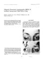
Show Joumal of Clinical Neuro- ophlhalmology 11( 1): 55- 56.1991. Tonic Pupil and Czarnecki's Sign Following Third Nerve Palsy Terry A. Cox, M. D., Robert A. Goldberg, M. D., and Jack Rootman, M. D. © 1991 Raven Press, Ltd., New York A 71- year- old woman developed abnormal pupillary function in one eye after a third nerve palsy. Stimulation with light caused segmental constriction of the pupil, the near reflex was normal, and gaze upward elicited constriction of portions of the sphincter that were unreactive to light. This combination of findings has not been reported previously. We believe that this case supports the idea that a tonic pupil can be caused by aberrant reinnervation of the ciliary ganglion. Key Words: Pupil- Reflex, pupillary- Oculomotor palsy- Tonic pupil. From the Department of Ophthalmology, University of British Columbia, Vancouver, B. C., Canada ( T. A. C., J. R.), and the Jules Stein Eye Institute, Los Angeles, California, U. S. A. ( RA. G.). Address correspondence and reprint requests to Dr. Terry A. Cox at Department of Ophthalmology, University of Utah Health Sciences Center, 50 N. Medical Drive, Salt Lake City, UT 84132, U. S. A. 55 In 1978, Czarnecki and Thompson ( 1) described segmental contractions of the iris sphincter associated with eye movements in 7 of 14 patients with acquired oculomotor synkinesis ( aberrant regeneration of the third nerve). In 1985, Coppeto et al. ( 2) described four patients with pupillary light- near dissociation following oculomotor nerve palsies. We report a woman with both of these findings affecting the same eye as the only residual evidence of a third nerve palsy caused by an arteriovenous fistula. CASE REPORT A 71- year- old woman noted mydriasis of the right pupil following an eye examination. One week later, she noted ptosis of the upper lid on that side. She also began to notice pain in and around the eye. During the next month the ptosis improved, but the mydriasis and intermittent pain persisted. Examination at that time revealed signs of a partial third nerve palsy on the right. There was a I- mm ptosis of the upper eyelid, with a 4mm reduction in levator function. Adduction of the eye was limited by 50%, depression by 20%, and elevation by 40%. The right pupil was 1 mm larger than the left in room light; the two pupils were nearly equal in darkness. A computed tomography ( CT) scan was normal. Six weeks later ( 11 weeks after onset of symptoms) she continued to have intermittent periocular pain on the right, mostly at night. Examination revealed no ptosis, normal levator function, and normal eye movements. The right pupil continued to be dilated, but examination at the slit lamp biomicroscope revealed a segmental light response and a diffuse near response. A 60° segment centered at the 9 o'clock position reacted well to light, and another 60° segment centered at the 3 0' clock 56 T. A. COX ET AL. REFERENCES out a red eye: an unusual presentation of dural arteriovenous shunts in the region of the cavernous sinus). It is possible that there was a preexisting tonic pupil, but we feel that all the pupillary findings were caused by the same lesion. Our patient has most of the pupillary signs associated with the tonic pupil of Adie's syndrome. She did not have noticeable tonic redilatation, but this sign is frequently absent in these cases. She also had segmental contraction of the pupillary sphincter with eye movement- a sign of oculomotor synkinesis after third nerve palsy. This combination of findings has not been reported previously. We believe that this case provides support for the hypothesis that a tonic pupil can result from aberrant reinnervation of the ciliary ganglion. The change in pupillary segments that reacted to light between the second and third exams could be explained by ongoing damage to preganglionic pupillary fibers with sporadic reinnervation; this mechanism has been proposed to explain primary aberrant regeneration of the oculomotor nerve caused by cavernous sinus masses ( 4). • Thirty minutes after pilocarpine 0.05% o. U, TABLE 1. Pupillary measurements 2.8 3.0 3.3 3.2 5.4 3.7 Left eye ( mm) 4,2 2.8 2.6 4.8 5.2 3.7 Right eye ( mm) Second exam Bright light Dim light Near Third exam Bright light Near Drops · position reacted less briskly. The remainder of the pupil failed to respond to light; however, the entire pupil reacted well to a near stimulus. Segmental reaction of the pupil with eye movements ( Czarnecki's sign) was not sought. Table 1 contains measurements of the pupil from photographs taken at this examination. The patient's intermittent ocular pain continued. Ten months after onset of her mydriasis, she first noticed a bruit synchronous with her heart beat. Examination 9 months later revealed signs of an arteriovenous fistula and a mild right sixth nerve palsy. There was a loud, high- pitched bruit centered over her right eye, but audible throughout the anterior cranium. There was a 3- mm proptosis on the right, and conjunctival and episcleral veins were dilated. There was no ptosis. Adduction, depression, and elevation of the eye were normal. Abduction was limited by about 20%. There continued to be pupillary light- near dissociation, but the pupillary response to light had changed. Twenty- degree segments centered at 1, 7, and 11 0' clock reacted well to light, and a smaller segment centered at the 4: 30 position reacted less well. The entire pupil reacted well to near, even when the eye was held in the primary position. The pupillary outline was irregular, and there was stromal streaming ( 3) along the inferior border. There was no segmental constriction of the pupil on gaze right or left; however, looking upward elicited brisk constriction of 20° pupillary segments centered at 8 and 10 o'clock in areas that did not react to light. Looking downward led to relaxation of these segments. Instillation of pilocarpine 0.05% led to more pupillary constriction on the right than on the left ( Table 1). Deep tendon reflexes were normal. DISCUSSION Our patient's third nerve palsy was almost certainly caused by the arteriovenous fistula. Two similar cases were presented at the Neuroophthalmology Congress, Vancouver, B. C., May 1988 ( Hawke SH, Mullie MA, Hoyt WF, Hallinan JM, Halmagyi GM: Painful third nerve palsy with- 1. Czarnecki JSc' Thompson HS. The iris sphincter in aberrant regeneration of the third nerve, Arch Ophthalmol 1978; 96: 1606- 10. 2. Coppeto JR, Monteiro MLR, Young D. Tonic pupils followmg oculomotor nerve palsies. Ann Ophthalmol 1985; 17: 585- 8. 3. Thompson HS. Segmental palsy of the iris sphincter in Adie's syndrome. Arch OphthalmoI1978; 96: 1615- 20. 4. Cox TA, Wurster JB, Godfrey WA. Primary aberrant oculomotor regeneration due to intracranial aneurysm. Arch Neural 1979; 36: 570- 1. ",' 1111 Neuro, ophthalmol, Vol. 11, No. 1, 1991 / |