| OCR Text |
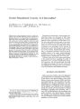
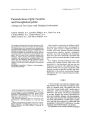
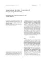
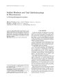
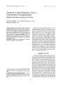
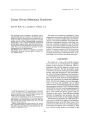
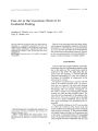
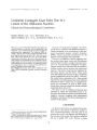
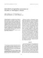
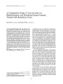
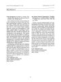
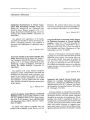
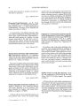
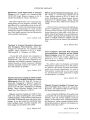
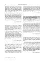
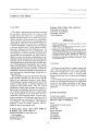
Show Journal of Cl,lllcal Nelao~ophthall1lology 13(j): 54-58, 1993. Unilateral Conjugate Gaze Palsy Due to a Lesion of the Abducens Nucleus Clinical and Neuroradiological Correlations Genjiro Hirose, M.D., Ph.D., Kei Furui, M.D., Akira Yoshioka, M.D., Ph.D., and Koichiro Sakai, M.D., ph.D. © 1993 Raven Press, Ltd 'Jew York We report a case of left-sided horizontal gaze palsy, ipsilateral adduction weakness, and left peripheral facial weakness, all of which indicate the lesion in the left median pontine tegmentum. The enhanced MRIs revealed a discrete left median pontine tegmental lesion, involving the abducens nucleus, MLF, and facial nerve knee. This lesion spared the area of the left PPRF. Among these structures, the area of the abducens nucleus seems to be responsible for the unilateral horizontal gaze palsy. We are not aware of any previous precise neuroradiological documentation of unilateral paralysis of conjugate gaze due to a lesion of the abducens nucleus by sagittal and horizontal MRIs. Key Words: Horizontal gaze palsy-Abducens Nucleus- MRI-Paramedian pontine reticular formation (PPRF). From the Department of Neurology, Kanazawa Medical University. Uchinada, Kahoku. Japan. Address correspondence and reprint requests to Dr. Genjiro Hirose. Department of Neurology. Kanazawa Medical University. Uchinada, Kahoku. Ishikawa Pref., Japan. 920-02. 54 Paralysis of horizontal conjugate eye movements seen in patients with brainstem lesions is usually attributed to the involvement of the ipsilateral paramedian pontine reticular formation (PPRF), which is generally considered to be the pontine horizontal gaze center clinically. However, considerable evidence suggested that the pontine lateral gaze center and the abducens nucleus might constitute a single anatomical structure (1,2). Axonal transport techniques and physiological studies have indicated that the abducens nucleus contains motor neurons and internuclear neurons. The former innervate the ipsilateral lateral rectus muscle, and the latter, whose axons cross the midline and ascend via the contralateral medial longitudinal fasciculus (MLF), innervate the medial rectus subnucleus (3,4). These studies indicate that ascending projections from abducens interneurons playa major role in conjugate horizontal eye movements. In spite of these experimental data, there have been few clinical examples supporting these data (5-8). We report a case of unilateral horizontal gaze paralysis in whom the responsible lesion is restricted to the abducens nucleus as documented by magnetic resonance imagings with enhancement techniques and discuss the clinicoradiological correlation in this patient. CASE REPORT An 80-year-old woman was admitted to the hospital with a I-day history of dizziness, double vision, and facial asymmetry. One day prior to admission she awoke with diplopia and drooping of the left side of her mouth. She could not close her left eye or gargle with water. Because of these HORIZONTAL GAZE PALSY 55 symptoms she was seen by a family doctor who referred her for further examinations. She denied other symptoms such as headache, nausea, or vomiting. She had been taking an oral antidiabetic drug with good control. On examination, she was alert and oriented to three spheres. Cognition was quite appropriate. Her speech was mildly dysarthric due to left facial weakness. Her pupils were isocoric and reacted normally. She had a tendency to look to the right side. She had a complete horizontal palsy of saccades and pursuit eye movements to the left beyond the midposition (Fig. 1). Oculocephalic maneuver did not elicit eye movements toward the left. With the attempt to look to the right, her right eye abducted fully with a coarse horizontal nystagmus, and her left eye adducted incompletely, indicating a left internuclear ophthalmoplegia. The return eye movement from the right extreme position to the midline was preserved with slow sac-cades. Convergence was present. She had left upper and lower facial weakness; taste was preserved. The rest of the cranial nerves were normal. She had no limb weakness. Pain and temperature sensation were normal in her limbs and her trunk. Her deep tendon reflexes were normal and no cerebellar signs were noted. The plantar response was equivocal on the right side. A left lower pontine medial tegmental lesion was diagnosed. Serial CT scans with enhancement were all reported negative. MRI 2 weeks after admission revealed a discrete high-signal lesion in the area of the medial lower pontine tegmentum on the left side by the T2 (TR:2,500, TE:90) images. This lesion was not noted by the II images (Fig. 2A,B). The lesion was enhanced by Gd-DTPA (Fig. 3A-C). This lesion gradually reduced in size on MRI 6 weeks later (Fig. 3D). These neuroradiological findings favor the diagnosis of ischemic infarction rather than hemorrhage or hemorrhagic infarction. FIG. 1. Extraocular movements of the patient on admission. Complete horizontal gaze palsy to the left beyond the midposition is noted with incomplete adduction of the left eye. Jelm N~uro-ol'hthalmol, Vol. 13, No.1, 1993 56 G. HIROSE ET AL. FIG. 2. Nonenhanced T1 images of the patient. Both (A) sagittal and (B) horizontal T1 images failed to show the responsible lesion in the pontine tegmentum. The left adduction weakness disappeared within 10 days after admission with a gradual recovery of the right adduction weakness next. At this stage she still had a left lateral rectus muscle weakness, and this cleared completely within about 2 months. Her left facial muscle weakness cleared last, after 3 months. AC and DC electro-oculographic (EGGs) recordings on hospital day 50 revealed incomplete slow saccadic eye movement toward to the left side and FIG. 3. Enhanced MRI findings of this patient. (A) Coronal view; a midline high-signal lesion is noted in the area of the lower pons on the 14th day of the illness. (B) Sagittal view; a high-signal lesion is located at the floor of the 4th ventricle at the level of the lower pons. (C) Horizontal view; a high-signal lesion is located close to the midline on the left medial tegmentum dorsal to PPRF, involving abducens nucleus, facial nerve knee, and MLF. (0) Horizontal view; 6 weeks later, a discrete enhanced lesion became smaller in size with clearing symptoms. JClin Neuro-ophthalmol. Vol. 13. No. L 1993 HORIZONTAL GAZE PALSY 57 saccadic eye movements toward the right in the eye tracking test. The optokinetic nystagmus (OKN) with quick phases to the patient's left was grossly defective when a drum rotated from left to right (Fig. 4). DISCUSSION Lesions in the abducens nucleus produce paralysis of the ipsilateral lateral rectus muscle and a paresis of contralateral ocular adduction on attempted horizontal gaze toward the side of the lesion. This symptomatology is quite different from the ipsilateral lateral rectus weakness secondary to the lesion in the abducens nerve fascicle, which is far more common. Carpenter and his associates (1) reported that the lesion strictly confined to the abducens nucleus showed an ipsilateral conjugate gaze palsy, involving the ipsilateral lateral rectus and contralateral medial rectus muscles in monkeys. Since then, considerable evidence suggests that the pontine center for lateral gaze and the abducens nucleus may constitute a single anatomical structure. Axonal transport techniques and physiological studies have indicated that the abducens nucleus contains motor neurons that innervate the lateral rectus muscle and internuclear neurons, which cross the midline, ascend via the contralat- ,."."""."""", " .. , .. ",.",., " ,' Right OKN eral MLF, and innervate the oculomotor nucleus (2-4). From these data, the paresis of contralateral ocular adduction, which is seen in the paralysis of lateral gaze and the paresis of ipsilateral adduction in anterior internuclear opthalmoplegia are now believed to result from the involvement of abducens internuclear neurons or their ascending axons. In our patient, neurological deficits consist of left-sided horizontal gaze paralysis, adduction weakness of the left eye and left-sided peripheral facial nerve palsy. These deficits were interpreted that this patient had left-sided gaze palsy and an internuclear ophthalmoplegia on the same side ("one and a half" syndrome) with the left peripheral facial nerve palsy. All of these symptoms indicate the lesion in the left median pontine tegmentum. This site of involvement was confirmed by the T2-weighted MRIs and gadoliniumenhanced MRls. The lesion seen on MRl involved the abducens nucleus, facial nerve knee, and MLF on the left side, explaining the patient's signs. There is a possibility that the PPRF might be also involved in our case. But PPRF is anatomically situated near the midline and ventrally to the MLF between the abducens and trochlear nuclei. It was not seen to be involved in the MRl of our patient (Fig. 3). Bronstein et a1. (8) reported MRl findings in seven patients with unilateral gaze palsy, but Left OKN SP Velocity ! 'Illl!!!!!'" ''''''!!III' , , I '" """,P,,,,,,,,.II'" !ILl I' ! I ., ,., ""••,_.,••.".,,,.'-.lW ...,,,, .. o 100 0 FIG. 4. Optokinetic nystagmus (OKN) recordings on, th~ 50th day of the illness. The notation Right OKN refers.to a stimulus which normally induce OKN to the patient s nght Side, actually the drum rotated around the patient With stripes passing from the right to the left. Left OKN ref~rs to the OKN, when stripes pass from the patient's left to the right. SP Velocity indicates the slow phase velocity, a~d an upward. and downward deflection of the EOGs denotes an eye movement to the right and the left, respectively. Calibrations are shown by vertical bars. JOin Neuro-ophtllQlmol, Vol. 13, No.1, 1993 58 G. HIROSE ET AL. only one of those had a localized lesion in the abducens nucleus secondary to a small hemorrhage. Even in this case, only the horizontal image was analyzed for localization. According to Pierrot-Deseilligy, horizontal gaze palsy secondary to the lesion in the paramedian pontine reticular formation, which he designates as the "Pontine reticular formation syndrome," is different from the syndrome due to the lesion in the abducens nucleus. In the PPRF syndrome, all ipsilateral saccades, including voluntary saccades and quick phases of nystagmus as well as the return phase of the contralateral hemifield eye movement are diminished. In our case, the return movement from the right abducted position to the midline was well preserved, supporting the MRI evidence that responsible lesion was in the abducens nucleus. As for the etiology of our case, her clinical symptoms and hospital course with complete clearing of the symptoms favor the diagnosis of ischemic cerebrovascular disease secondary to the occlusion of the paramedian perforating artery of the pons. REFERENCES 1. Carpenter MB, McMaster RE, Hanna GR. Disturbances of conjugate horizontal eye movements in the monkey. I. I Cli" Neuro-ophthalmol, Vol. 13, No. 1. 1993 Physiological effects and anatomical degeneration resulting from lesions of the abducens nucleus and nerve. Arch NeuroI1963; 8:231-47. 2. Graybiel AM, Hartwieg EA. Some afferent ways in the cat and rhesus monkey. In: Baker R, Berthoz A, eds. Control of gaze by brain stem neurons. Developments in neuroscience. Amsterdam: ElsevierfNorth-Holland, 1974:79--88. 3. Graybiel AM, Hartwieg EA. Some afferent connections of the oculomotor complex in the cat: an experimental study with tracer techniques. Brain Res 1974;81:543-51. 4. Baker R, Highstein SM. Physiological identification of interneurons and motoneurons in the abducens nucleus. Brain Res 1975;91:292--8. 5. Bennett H, Savill TH. A case of permanent conjugate deviation of the eyes and head, the result of a lesion limited to the sixth nucleus; with remarks on associated lateral movements of the eyeballs, and rotation of the head and neck. Brain 1889;12:102-16. 6. Meienberg 0, Buttner-Ennever JA, Kraus-Ruppert R. Unilateral paralysis of conjugate gaze due to lesion of the abducens nucleus: clinico-pathological case report. Neuroophthalmology 1981;2:47-52. 7. Pierrot-Deseilligny CH, Goasguen J. Isolated abducens nucleus damage due to histiocytosis X. Brain 1984;107:101932. 8. Bronstein AM, Rudge P, Gresty MA, Du Boulay G, Morris J. Abnormalities of horizontal gaze: clinical, oculographic and magnetic resonance imaging findings. II. Gaze palsy and internuclear ophthalmoplegia. JNeurol Neurosurg Psychiatry 1990;53:200-7. 9. Pierrot-Deseilligy C, Chain F, Lherrnitte F. Syndrome de la formation reticula ire pontique: precisions physiopathologiques sur les anomalies des mouvements oculaires voluntaires. Rev Neurol 1982;138:517-32. |