| OCR Text |
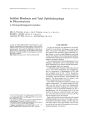
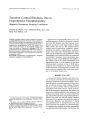
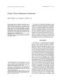
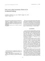
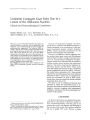
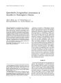
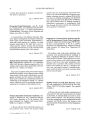
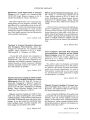
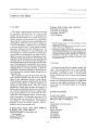
Show ]ounlal of Clinical Ncuro-ophth111molosy 13(1)· ~1-~(I. 1993 Visual Loss as the Initial Presentation of Nasopharyngeal Carcinoma Ling-Yuh Kao, M.D., Huei-Chun Chuang, M.D., and Yu-Song Liang, M.D. © 1993 Raven Press, UO"' York Eye symptoms and cranial nerve involvement are rather common in nasopharyngeal carcinomas, but early invasion of the optic nerve is very rare. Two cases of nasopharyngeal carcinoma that presented initially with visualloss are reported. Key Words: Nasopharyngeal carcinoma-Optic nerveCT scan-Orbital apex compression. From the Department of Ophthalmology. Chang Gung Memorial Hospital, Chang Gung Medical College. Taipei. Taiwan, Address correspondence and reprint requests to Dr. L. Y, Kao at Department of Ophthalmology. Chang Gung Memorial Hospital, 199 Tun Hwa North Road. Taipei. Taiwan. 24 Nasopharyngeal carcinoma is a very common malignant disease among the Chinese, especially among the inhabitants of the southeast provinces of China and Taiwan. Based on surgical biopsy specimens, nasopharyngeal carcinoma is the most common cancer in males and the third most common in females in Taiwan (1). The diagnosis of nasopharyngeal carcinoma sometimes can be a challenge to physicians. This is because nasopharyngeal neoplasms sometimes hide all nasal symptoms and present nonspecific signs such as diplopia, hearing loss, or facial pain as the first manifestation. Nasopharyngeal cancers remain one of the most easily misdiagnosed malignant diseases. We recently encountered two patients who experienced painless monocular blindness as the initial symptom of nasopharyngeal carcinoma. CASE REPORT Case 1 A 68-year-old male was seen in May 1989 with the chief complaint of progressive right eye visual loss of 3 months duration. Past history revealed that he had suffered from hearing loss in the right ear for 10 years because of chronic otitis media. Eye examination revealed that the vision was no light perception in his right eye, and was corrected to 20120 in the left eye. The right eye showed an amaurotic pupillary response, but the eye movements and ocular fundi were all normal. Computed tomography (CT) scan demonstrated a space-occupying lesion extending from the right orbital apex to the parasellar area (Fig. 1). A blind biopsy from the nasopharynx was done, and pathologic examination of the specimen revealed "nonkeratinizing squamous cell carcinoma." The patient received radiotherapy thereafter, but 1 year after completing the radiotherapy, pro- NPC PRESENTED AS VISUAL LOSS 25 FIG. 1. CT brain scan of Case 1 showing a spaceoccupying lesion extended from right orbital apex to parasellar area. ptosis and disturbed ocular motility appeared, and orbital invasion by the nasopharyngeal carcinoma became obvious. Case 2 A 54-year-old male was seen initially in December 1985 at a retina clinic with the chief complaint of progressive right eye blurred vision of one month's duration. Examination revealed a corrected visual acuity of 20/200 in the right eye, and 20/20 in the left eye. The eye movements were normal. Fundus examination disclosed normal optic discs, but some pigmentary mottling was seen at the posterior pole of the right eye. Fluorescein angiography demonstrated some window defects superior to the macula. Under the impression of central serous choroidoretinopathy, a focal retinal photocoagulation was done. However, the visual impairment became worse, and by March 1986 the visual acuity was no light perception in the right eye. The patient did not return until May 1986, when he presented with a blind right eye but with additional signs. Ptosis of the right eye, limitation of eye movement in all directions, and numbness of the right face indicated multiple cranial nerve involvement. A CT scan demonstrated a space-occupying lesion situated at paracavernous sinus and orbital apex area (Fig. 2). Patient was referred to the neurosurgical department and a craniotomy was performed. The tumor mass was removed and the specimen disclosed a "poorly differentiated carcinoma." The orbital and other cranial nerve involvements appeared 5 months after the initial presentation of visual loss, and there were no accompanying nasal symptoms during the whole course of the illness. DISCUSSION The common clinical manifestations of nasopharyngeal carcinoma are neck mass, blood-tinged sputum or rhinorrhea, hearing loss, headache, and cranial nerve palsy (2). Cranial nerve involvement was observed in 20-25% of nasopharyngeal carcinoma patients at the first examination (3,4). The 5th cranial nerve was the most commonly affected, followed by the 6th cranial nerve. Isolated optic nerve involvement was rarely reported as the initial manifestation (5,6), but has been frequently found late in the course, and usually associated with extraocular muscle weakness. The optic nerve encasement by tumor was well demonstrated by computerized cranial tomography. Orbital apex in- FIG. 2. CT brain scan of Case 2 revealed a spaceoccupying lesion situated at orbital apex and paracavernous sinus area. 1Clill Nelln>-o""'h"I",,,I. Vol. 13. No. l. 1993 26 L- Y. KAO ET AL. vasion with paracavernous sinus extension without bony destruction was disclosed in these two cases, indicating probably that the lesion went through the foramen lacerum, with the optic nerve invasion (7). The reason why these two cases showed optic nerve involvement first and spared the extraocular muscles at onset remained puzzling. SUMMARY Visual loss as the initial presentation of nasopharyngeal carcinoma is very unusual. When a patient experiences progressive visual loss of unknown cause, a detailed nasopharynx and neuroimaging study is indicated, especially in areas of high prevalence. I Clin Neuro-ophthalmol, Vol. 13, No.1, 1993 REFERENCES 1. Yeh S. Some geographic aspects of most common diseases in Taiwan: part two. Int Pathal 1966;7:24-8. 2. Mow-Ming Hsu, Shih-Mien Tu. Nasopharyngeal carcinoma in Taiwan: clinical manifestation and results of therapy. Cancer 1983;52:362-8. 3. Chen MS, Lin FJ, Tang SG, Leung WM, Leung Wan: clinical significance of cranial nerve deficit in the therapy of nasopharyngeal carcinoma. Br I Radial 1989;62:739--43. 4. Huang SC Nasopharyngeal carcinoma, a review of 1605 patients treated radically with Co60. Int I Radiat Oneal Bioi Phys 1980;6:401-7. 5. Chen C Early invasion of the optic nerve by nasopharyngeal carcinoma: a report of 2 cases. Chung Hua Yen Ka Tsa Chih 1980;16(4):372. 6. Carlin L. Monocular blindness in nasopharyngeal cancer. Arch Neural 1981;38:600. 7. Huang KM, Hsu JC The coronal section study of computed tomography in the intracranial invasion by nasopharyngeal carcinoma. Chin I Radial 1982;7:177-94. |