| OCR Text |
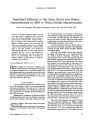
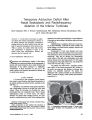
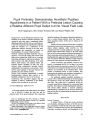
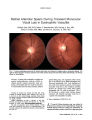
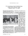
Show ORIGINAL CONTRIBUTION Pupil Perimetry Demonstrates Hemifield Pupillary Hypokinesia in a Patient With a Pretectal Lesion Causing a Relative Afferent Pupil Defect but No Visual Field Loss Eleni Papageorgiou, MD, Thomas Wermund, MD, and Helmut Wilhelm, MD Background: Lesions affecting the pretectum or the brachium of the superior colliculus (brachium) and sparing the optic tract cause a contralateral relative afferent pupil defect (RAPD) but no visual field loss. It has been assumed that the pupillomotor pathways within the brachium are a continuation of the pupillomotor pathways traveling in the optic tract. To investigate this assumption, we looked for hemihypokinesia by means of pupil perimetry. Methods: Pupillary hemifield stimulation was per-formed in a 65-year-old woman with normal visual fields and an isolated left RAPD due to a cerebral hemorrhage affecting the right dorsal midbrain. The pupil responses from light stimulation of the nasal inferior, nasal superior, and temporal inferior and temporal superior quadrants of both eyes were recorded using computerized binocular infrared pupillography. Each stimulus was presented 5 times and the mean amplitude of the pupil response was calculated for each stimulus location. Results: Pupil perimetry demonstrated a marked hemihypokinesia (reduced light reaction) in the hemifield contralateral to the site of the lesion. Conclusions: Our experiment suggests that the brachium is indeed a continuation of the afferent pupillary fibers traveling in the optic tract. (J Neuro-Ophthalmol 2009;29:33-36) n the human pupillary light reflex pathway, the afferent arc is believed to connect the optic tract and the pretectal nuclei of the dorsal midbrain. This connection, called the brachium of the superior colliculus (brachium), is said to contain afferent pupillary fibers that bypass the lateral geniculate nucleus (LGN) and are relayed to the midbrain (1). Patients with total optic tract lesions show a complete contralateral homonymous hemianopia and a contralateral relative afferent pupillary defect (RAPD) (2). The contralateral RAPD is usually attributed to the larger size and the greater photoreceptor density of the nasal retinal field, as well as to the asymmetrical chiasmal decussation of fibers arising from nasal and temporal retina (3-5). Infrared pupillography has suggested that the contralateral RAPD in optic tract lesions reflects the difference in light sensitivity between the intact temporal and nasal hemiretinas (6). Lesions restricted to the brachium, which are rare, result in a contralateral RAPD (2), but there is no visual field defect because the optic tract is spared (3,5). The aim of this study was to determine whether a lesion in the dorsal midbrain, the termination of the brachium fibers, would produce a hemianopic defect by pupil perimetry, thus implying that the brachium truly contains a continuation of the pupillomotor fibers traveling in the optic tract. We performed pupillary hemifield stimulation in a patient with a unilateral hemorrhage in the basal ganglia that appeared to involve the dorsal midbrain but not the optic tract. This patient had a contralateral RAPD but a normal visual field as determined by conventional light perimetry. The finding of a similar pupillographic pattern in both clinical entities indicates a common underlying pathophysiologic mechanism for the observed RAPD and thus provide clues for understanding the architecture of the pupillary pathway. METHODS Patient Features In January 2006, a 65-year-old woman developed acute headache, dysphagia, heaviness of the left arm and leg, and numbness on the left side of the body. There was a history of increased blood pressure and hyperlipidemia. Neurologic examination showed mild hemiparesis of the Center for Ophthalmology (PE, HW), University of Tu¨bingen, Tu¨bingen, Germany; and Helios Kliniken Schwerin (TW), Department of Ophthalmology, Schwerin, Germany. This work was supported by the European Union (PERACT-Marie Curie Early Stage Training MEST-CT-2004-504321). Address correspondence to Eleni Papageorgiou, Centre for Ophthal-mology, University of Tu¨bingen, Schleichstrasse 12-16, 72076, Tu¨bingen, Germany, E-mail: e_papage@yahoo.com J Neuro-Ophthalmol, Vol. 29, No. 1, 2009 33 J Neuro-Ophthalmol, Vol. 29, No. 1, 2009 Papageorgiou et al FIG. 1. Ascending axial CT sections (left to right) show increased attenuation in the right dorsal midbrain, right basal ganglia, right posterior thalamus, and third ventricle, but sparing the optic tract. This abnormality is consistent with intraparenchymal and intraventricular hemorrhage. left arm and leg, a left hemisensory deficit, and a right gaze palsy. Cranial CT demonstrated a hemorrhage in the right basal ganglia, which also involved the right posterior thalamus and the right dorsal midbrain (Fig. 1). It appeared to spare the right optic tract. Seven months after clinical onset, all of the patient's neurologic manifestations had resolved except a left hemi-sensory deficit. She did not report any visual difficulties. Visual acuity was 20/20 in both eyes with normal color vision and full visual fields as assessed with Octopus 101 static automated perimetry. Results of slit-lamp and ophthalmoscopic examinations were unremarkable, and ocular motility was normal in both eyes. The pupils were of equal size, but there was a left RAPD of 0.9 log unit by neutral density filters and 1.33 log units by an automated pupillographic swinging flashlight test. Pupil Perimetry We performed pupil perimetry using light stimuli (circles) of 12 diameter and 8 cd/m2 luminance presented on a 19-inch computer monitor under mesopic conditions (background luminance of 1 cd/m2). A custom-built proto-type was used to provide the hemifield stimuli on the monitor and to record simultaneously the right and left horizontal pupil diameters at 25 times per second. All parameters concerning pupil perimetry are listed in Table 1. We used one stimulus in each quadrant (Fig. 2). Each stimulus was presented 5 times, and the mean amplitude of the pupil response was calculated for each stimulus location using computer software. In accordance with the methods of Kardon et al (6), the term ‘‘hemifield'' is used to refer to the corresponding visual field side. RESULTS Based on the mean amplitudes of the pupil responses in each quadrant (Table 2), a pupillary hemihypokinesia was present with loss of pupillomotor sensitivity contra-lateral to the side of the lesion. The mean amplitudes of the pupil responses in the left hemifields were very low in both eyes, reflecting a homonymous pattern of pupillomotor sensitivity loss contralateral to the optic tract lesion (Fig. 2). The pupil response from the functioning temporal hemifield ipsilateral to the tract lesion (right eye) was greater than that from the functioning nasal hemifield contralateral to the tract lesion (left eye). DISCUSSION The pupil light reflex in isolated pretectal lesions provides a unique model for selectively studying the pupillary pathway anatomy because the afferent pupillo-motor fibers here are anatomically separate from the visual fibers. The clinical findings in the present patient are consistent with a lesion that selectively damaged the pupillary afferents in the brachium or its destination in the pretectal nucleus. Although our patient had a relatively large hemorrhage, the absence of a homonymous field defect excludes a substantial lesion of the optic tract. In addition, the normal color of the optic disc proves that the optic tract is not involved. TABLE 1. Instrumentation and stimulus characteristics used in pupil perimetry Campimetry monitor Testing distance Visual field covered Stimulus size Stimulus luminance Pupillography Data analysis Belinea, model 106020, 19-inch 20 cm 36 27 Adjustable, here 12 8 cd/m2 Customized monocular infrared video, 25 Hz Adjustable artefact management, repetition of insufficient pupillograms, determination of constriction amplitude 34 q 2009 Lippincott Williams & Wilkins Pupil Perimetry J Neuro-Ophthalmol, Vol. 29, No. 1, 2009 FIG. 2. Pupil perimetry results. The black circle between 10 and 20 in each quadrant indicates the position of the presented stimuli (12 diameter, 8 cd/m2 brightness). Note the marked hemihypokinesia in the left hemifield of both eyes as well as the difference in pupil response between the functioning (temporal) hemifield of the right eye and that of the left eye (nasal), which represents the intereye difference observed clinically (left RAPD). Lesions confined to this anatomic region are rare and may often remain undetected, as they are not accompanied by a visual deficit. Behr (7) first described a RAPD in two patients with no detectable visual dysfunction after stroke. He proposed that the lesion involved the afferent pupillary pathway in the brachium or pretectum (7). Nine such cases indicating affection of the brachium or the pretectum were subsequently published between 1984 and 2005 (2-5,8-12). The majority of the reported patients manifested additional neurological signs and accompanying ocular motility disorders, as did our patient. The underlying pathologic lesion in the reported cases was tumor, hemorrhage, or infarction in the dorsal midbrain and the RAPD was, as in our patient, always contralateral to the lesion (2-5,7-12). TABLE 2. Contraction amplitude values of the pupil response in each of the four quadrants Left Eye Right Eye Mean Mean amplitude amplitude (mm) SD (mm) SD Inferior temporal Inferior nasal Superior nasal Superior temporal 0.18 0.62 0.42 0.18 0.03 0.04 0.06 0.06 1.20 0.50 0.41 1.17 0.04 0.06 0.10 0.17 The pupillary hemihypokinesia we found in our patient represents the pupillographic correlate of the clinically observed contralateral RAPD. These results are identical to those we would expect in an optic tract lesion. Consequently, the RAPD in isolated unilateral pretectal lesions has the same origin as the RAPD in optic tract lesions, which primarily represents a difference in light sensitivity between the intact nasal and temporal hemi-retinas (6). Even in retrogeniculate lesions, a contralateral RAPD may be observed (13,14). There are strong hints that even in those cases the lesion involves the brachium of the dorsal midbrain. Our findings suggest that there is no need to posit a cortico-pretectal pathway to account for the RAPD in brachium or pretectal lesions, which may play a role in other pupillary phenomena (15). REFERENCES 1. Loewenfeld IE. The Pupil: Anatomy, Physiology, and Clinical Appli-cations, Vol 1. Boston: Butterworth and Heinemann; 1999:203-28. 2. Forman S, Behrens MM, Odel JG, et al. Relative afferent pupillary defect with normal visual function. Arch Ophthalmol 1990;108: 1074-5. 3. Chen CJ, Scheufele M, Sheth M, et al. Isolated relative afferent pupillary defect secondary to contralateral midbrain compression. Arch Neurol 2004;61:1451-3. 4. Taguchi H, Kashii S, Kikuchi M, et al. Superior oblique paresis with contralateral relative afferent pupillary defect. Graefes Arch Clin Exp Ophthalmol 2000;238:927. 5. Eliott D, Cunningham ET, Miller NR. Fourth nerve paresis and ipsilateral relative afferent pupillary defect without visual sensory 35 J Neuro-Ophthalmol, Vol. 29, No. 1, 2009 Papageorgiou et al disturbance. A sign of contralateral dorsal midbrain disease. J Clin Neuroophthalmol 1991;11:169-72. 6. Kardon R, Kawasaki A, Miller NR. Origin of the relative afferent pupillary defect in optic tract lesions. Ophthalmology 2006;113: 1345-53. 7. Behr C. Hemianopische Pupillenstarre ohne Hemianopsie. Z Prakt Augenheilkd 1926;58:398-406. 8. King JT Jr, Galetta SL, Flamm ES. Relative afferent pupillary defect with normal vision in a glial brainstem tumor. Neurology 1991;41:945-6. 9. Ellis CJ. Afferent pupillary defect in pineal region tumour. J Neurol Neurosurg Psychiatry 1984;47:739-41. 10. Johnson RE, Bell RA. Relative afferent pupillary defect in a lesion of the pretectal afferent pupillary pathway. Can J Ophthalmol 1987;22: 282-4. 11. Girkin CA, Perry JD, Miller NR. A relative afferent pupillary defect without any visual sensory deficit. Arch Ophthalmol 1998;116:1544-5. 12. Staubach F, Pieh C, Maier P, et al. Relative afferent pupillary defect with normal vision and vertical strabismus-implications for pupillary pathway anatomy. Graefes Arch Clin Exp Ophthalmol 2007;245: 321-3. 13. Wilhelm H, Wilhelm B, Petersen D, et al. Relative afferent pupillary defects in patients with geniculate and retrogeniculate lesions. Neuroophthalmology 1996;16:219-24. 14. Papageorgiou E, Ticini LF, Hardiess G, et al. The pupillary light reflex pathway: Cytoarchitectonic probabilistic maps in hemianopic patients. Neurology 2008;70:956-63. 15. Wilhelm BJ, Wilhelm H, Moro S, et al. Pupil response components: studies in patients with Parinaud's syndrome. Brain 2002;125:2296-307. 36 q 2009 Lippincott Williams & Wilkins |