| OCR Text |
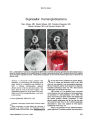
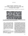
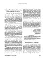
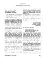
Show ORIGINAL CONTRIBUTION Anatomic Characteristics of the Ophthalmic and Posterior Ciliary Arteries Senem Erdogmus, MD and Figen Govsa, MD Background: There is little documentation of the course and relations of the ophthalmic artery (OA) and posterior ciliary arteries (PCAs). Methods: The anatomic characteristics of the OA and PCAs were determined from a dissection of 19 neoprene injected cadaver heads. Results: The intraorbital OA had three segments, considering its relation to the optic nerve in the sagittal plane. The first segment extended from the point where the OA entered the orbit to its curving point. The second segment coursed superomedially from the inferolateral part of the optic nerve, crossing the optic nerve either superiorly or inferiorly. The third segment extended from the curving point of the superomedial distal portion of the second segment to the vessel's termination. The OA was deviated at the junction of its first and second segments, defined as its ‘‘angle''; and at the junction of the second and third segments, defined as its ‘‘bend.'' The PCAs arose from the first OA segment, the angle of the OA, the second OA segment and the OA bend. The patterns of branching of the PCAs were medial and lateral and medial, lateral, and superior. The superior PCA and the lateral PCA arose mainly from the angle of the OA, whereas the medial PCA arose from the curving point of the OA. The most frequently observed PCA pattern was a medial PCA and a lateral PCA. The average diameters of the medial PCA, the superior PCA, and the lateral PCA were 0.65, 0.48, and 0.68 mm, respectively. In all cases, pial arteries branching from the PCA and supplying the optic sheath were observed to form a vascular plexus on the optic sheath. The OA and PCAs were surrounded by a network of sympathetic nerves. Conclusions: Because the most common pattern of PCAs is a medial and lateral branch, a surgical approach to the orbit from those directions carries a higher risk of damage to those vessels than a superior or inferior approach. (J Neuro Ophthalmol 2008;28:320-324) The superficial layers of the optic nerve head are supplied by the central retinal artery, and the deep layers are supplied by the posterior ciliary arteries (PCAs), branches of the ophthalmic artery (OA) (1-4). Ischemic disorders of the optic nerve head constitute an important cause of visual loss (3-7), spurring a search for methods to reliably evaluate the circulation to this tissue (3,4,7-9). Moreover, we have observed that the PCAs have a high risk of being damaged during surgery of the orbit. The aim of this study was, therefore, to investigate the precise anatomic characteristics of the OAs and PCAs. METHODS Dissection was performed on 19 adult male human cadavers (38 orbits), fixed with 10% formalin, in the Department of Anatomy, Faculty of Medicine, Ege University, Izmir, Turkey. After the skulls were opened and the brains were removed, a liquid latex 601 neoprene mixture colored with powder eosin paint was injected through the internal carotid artery. The orbital section of the frontal bone was removed by careful dissection, thus enabling the visualization of the orbital structures. A high-speed drill was used to remove the bony walls of the optic canal. After the removal of the bony walls and the connective fatty tissue of the orbit, the origin, position, branches, course, and anatomic relations of the PCAs were noted. Measurements in millimeters were made by means of a digital calliper. Statistical analyses were performed using SPSS version 10.0. Department of Anatomy, Faculty of Medicine, Ege University, Izmir, Turkey. Address correspondence to Prof. Dr. Figen Govsa, MD, Ege U¨ niversitesi Tıp Faku¨ltesi Anatomi Anabilim Dalı, TR 35100, Izmir, Turkey; E mail:figen.govsa@ege.edu.tr RESULTS The Intraorbital OA The course of the intraorbital part of the OA was macroscopically studied in three segments, considering its 320 J Neuro Ophthalmol, Vol. 28, No. 4, 2008 Posterior Ciliary Arteries J Neuro-Ophthalmol, Vol. 28, No. 4, 2008 FIG. 1. The posterior ciliary arteries (PCAs) in relation to the optic nerve and globe. 1, ophthalmic artery; 2, medial posterior ciliary artery (PCA); 3, lateral PCA; 4, superior PCA; 5, optic nerve; 6, globe; and 7, sympathetic nerves. FIG. 2. The lateral posterior ciliary artery (PCA) originating from the angle part of the ophthalmic artery. 1, ophthalmic artery; 2, nasociliary nerve; 3, lateral posterior ciliary arteries (PCA); 4, optic nerve; 5, globe; 6, superior oblique artery; 7, sympathetic nerves; and 8, pial artery. FIG. 3. The superior posterior ciliary artery (PCA) superior to the optic nerve. 1, superior PCA; 2, vascular network; and 3, sympathetic nerves. 321 J Neuro-Ophthalmol, Vol. 28, No. 4, 2008 Erdogmus and Govsa relation to the optic nerve in the sagittal plane. The first segment extended from the point where the OA entered the orbit to its curving point. This first segment of the OA lay very close to the optic nerve, free of orbital fat. The OA usually ran along the inferolateral aspect of the optic nerve. The second segment of the OA began where the artery approached the lateral side of the optic nerve and started to cross over it at a right, acute, or obtuse angle. The point at which the artery changed direction and shape represented the end of the second segment. This second segment coursed superomedially from the inferolateral optic nerve, crossing the optic nerve either superiorly or inferiorly. In the third segment of the intraorbital OA, the vessel was located on the medial part of the optic nerve and globe, as well as the lateral part of the superior oblique and the medial rectus, reaching the medial wall of the orbit close to the anterior ethmoid foramen. The OA deviated at two points: at the junction of its first and second segments, defined as the ‘‘angle,'' and at the junction of its second and third segments, defined as the ‘‘bend'' (1,2). The OA angle was observed to be obtuse; the OA bend was not as well defined. The PCAs The PCAs arose independently from the first OA segment, the OA angle, the second OA segment, and the OA bend. The PCAs coursed distally, dividing into multiple branches and piercing the sclera close to the optic nerve medially, laterally, and superiorly (Figs. 1-3). The branch-ing formations of the PCA are given in Table 1. A medial PCA stemmed from various origins such as the first OA segment, the OA angle, the OA bend, and the third OA segment. The size of the outer diameter of the medial PCA is indicated in Table 2. The origin of the medial PCA is indicated in Table 3. The lateral PCA arose from various origins, including the first OA segment, the OA angle, and the second OA segment, always lateral to the optic nerve (Fig. 1). The size of the outer diameter of the lateral PCA is given in Table 2. TABLE 1. Number and percentage of the posterior ciliary arteries Branching Pattern Medial 1 lateral Medial 1 lateral 1 superior Data are n (%). Right 12 (63.15) 7 (36.8) Left Bilaterally 14 (73.7) 11 (57.9) 5 (26.3) 4 (21.05) The descriptive anatomy of the lateral PCA is given in Table 4. Despite its various origins, including the OA angle, the second OA segment, and the OA bend, the superior PCA was always located superior to the optic nerve (Figs. 1 and 3). The size of the outer diameter of the superior PCA is given in Table 2. The descriptive anatomy of the superior PCA is summarized in Table 5. In all cases, the intraorbital part of the OA and the PCA gave off thin pial arteries that penetrated the superior surface of the optic sheath at a right angle. These arteries formed a subpial meshwork. This network contributed to the vascularization of the optic nerve fibers (Figs. 2 and 3). Also in all cases, the OA and PCAs were accompanied by fine nerve filaments and a plexus that seemed to arise from the carotid sympathetic nerves. These nerves surrounded and formed a network on the optic nerve sheath (Figs. 1-3). DISCUSSION Ophthalmic surgeons and neurosurgeons who oper-ate within the confines of the orbit must know the neurovascular anatomy of the optic canal and, in particular, the course of the PCAs. This knowledge is especially relevant in the management of clinoidal meningiomas with intraorbital extension, aneurysms, hematomas, and optic sheath meningiomas with intracranial extension and as visualization of the operative field is restricted by orbital fat in strabismus surgery (10-12). Moreover, the origin and branching pattern of the PCAs cannot be defined on preoperative neuroimaging studies because of their small caliber. Surgical damage to the PCA circulation can result in a variety of ocular and optic nerve vascular disorders, causing varying degrees of visual loss (7). However, there have been few investigations discussing the detailed micro-surgical anatomy of the PCAs. TABLE 2. Outer diameter of the posterior ciliary arteries Posterior ciliary artery Medial Lateral Superior Diameter Right 0.65 6 0.12 (0.43-0.97) 0.68 6 0.11 (0.51-0.82) 0.48 6 0.11 (0.34-0.65) Data are means (minimum-maximum). (mm) Left 0.65 6 0.14 (0.41-0.90) 0.67 6 0.14 (0.43-0.78) 0.54 6 0.09 (0.42-0.67) 322 q 2008 Lippincott Williams & Wilkins Posterior Ciliary Arteries J Neuro-Ophthalmol, Vol. 28, No. 4, 2008 TABLE 3. Number and percentage of the origin of the medial posterior ciliary artery arising from the ophthalmic artery Location of ophthalmic artery (OA) crossing of optic nerve Top Bottom Right Left Right Left Site of origin First OA OA angle Second OA OA bend Third OA n % n % n % n % 2 10.52 3 15.78 - - - - 5 26.31 3 15.78 - - - - 7 36.84 9 47.36 3 15.78 1 5.26 - - 1 5.26 2 10.52 2 10.52 Previous authors describe the origins of the PCAs as variable (13-16). Some authors stated that the PCAs arose from the first part of the OA, others claimed that they arose superior to the optic nerve in the second part, and still others reported that they arose medial to the optic nerve (6,7,8,13-16). In this study, the superior PCA and the lateral PCA most frequently branched from the angle part of the OA, whereas the medial PCA originated from its bend part. Our results are most compatible with those of Hayreh (7), Ettl et al (9), and in particular Onda et al (8). Sudakevitch (15) reported that the medial PCA, in common with the central retinal artery, was usually the first branch of the OA, that the lateral PCA was the second, and that both PCAs arose before the lacrimal artery, which was situated at the OA angle. As opposed to Sudakevitch, we have observed the medial PCA to arise independently from the OA. In their studies, previous authors have found that an eye might be supplied by one (in 3%-7%), two (in 25%- 48%), three (in 39%-50%), four (in 8%-17%), or five (in 2%-8%) PCAs arising from the OA (13-16). The most frequently observed branching pattern was the one for which there were one medial and one lateral PCAs. This study was in agreement with the observations in previous studies (1,7,9) with regard to the existence of two PCA branches in most cases. However, in less than one third of the cases, there were three branch PCA, a finding that differs markedly from those of Lang and Kageyama (16) and Ducournau (17). Previous authors have reported the intraorbital presence of the autonomic fibers arising from the internal carotid plexus (10,18). We have observed this neural network to arise from the intracranial sympathetic nerve fibers on the internal carotid artery and accompany the OA and PCAs, forming a network on the optic sheath. These fibers are thought to control blood supply to the intraorbital tissues and should be avoided during surgery (10,18,19). TABLE 4. Number and percentage of the origin of the lateral posterior ciliary arteries arising from the ophthalmic artery Location of ophthalmic artery (OA) crossing of optic nerve Top Bottom Right Left Right Left Site of origin First OA OA angle Second OA OA bend Third OA n % n % n % n % 3 11 - - 15.78 57.89 - - 4 12 - - 21.05 63.15 - - 3 1 1 - 15.78 5.26 5.26 - 2 1 - - 10.52 5.26 - - 323 J Neuro-Ophthalmol, Vol. 28, No. 4, 2008 Erdogmus and Govsa TABLE 5. Number and percentage of origin of the superior posterior ciliary arteries arising from the ophthalmic artery Location of ophthalmic artery (OA) crossing of optic nerve Top Bottom Right Left Right Left SiteofOrigin n % n % n % n % FirstOA - - - - - - - - OA angle 3 50.0 2 40.0 - - - - SecondOA 1 16.66 - - 1 16.66 - - OA bend 1 16.66 2 40.0 - - 1 20.0 ThirdOA - - - - - - - - REFERENCES 1. Hayreh SS. The ophthalmic artery. III. Branches. Br J Ophthalmol 1962;46:212-46. 2. Hayreh SS, Dass R. The ophthalmic artery. II. Intra-orbital course. Br J Ophthalmol 1962;46:165-85. 3. Hayreh SS. Blood supply of the optic nerve head and its role in optic atrophy, glaucoma and oedema of the optic disc. Br J Ophthalmol 1969;53:721-48. 4. Hayreh SS. Pathogenesis of visual field defects: role of the ciliary circulation. Br J Ophthalmol 1970;54:289-311. 5. Hayreh SS. Blood supply of the optic nerve head. Ophthalmologica 1996;210:285-95. 6. van Overbeeke J, Sekhar L. Microanatomy of the blood supply to the optic nerve. Orbit 2003;22:81-8. 7. Hayreh SS. Posterior ciliary artery circulation in health and disease: the Weisenfeld Lecture. Invest Ophthalmol Vis Sci 2004; 45:749-57. 8. Onda E, Cioffi AG, Bacon DR, et al. Microvasculature of the human optic nerve. Am J Ophthalmol 1995;120:92-102. 9. Ettl A, Kramer J, Daxer A, et al. High resolution magnetic resonance imaging of neurovascular orbital anatomy. Ophthalmology 1997;104: 869-77. 10. Tsutsumi S, Rhoton AL Jr. Microsurgical anatomy of the central retinal artery. Neurosurgery 2006;59:870-9. 11. Erdogmus S, Govsa F, Celik S. Innervation features of the extraocular muscles. J Craniofac Surg 2007;18:1439-46. 12. Erdogmus S, Govsa F. Arterial vascularization of the extraocular muscles on its importance for orbital approaches. J Craniofac Surg 2007;18:1125-32. 13. Jimenez-Castellanos J, Carmona A, Castellanos L, et al. Microsurgical anatomy of the human ophthalmic artery: a mesoscopic study of its ori-gin, course and collateral branches. Surg Radiol Anat 1995;17:139-43. 14. Jo-Osvatic A, Basic N, Jukic T, et al. Topoanatomic relations of the ophthalmic artery viewed in four layers. Surg Radiol Anat 1999;21: 371-5. 15. Sudakevitch T. The variations in system of trunks of the posterior ciliary arteries. Br J Ophthalmol 1947;31:738-60. 16. Lang J, Kageyama I. The ophthalmic artery and its branches, measurements and clinical importance. Surg Radiol Anat 1990;12: 83-90. 17. Ducournau D. Systematisation Vasculaire de la Choroide. Lyon, France: Association Corporative des Etudiants en Medecine de Lyon; 1979:17-22. 18. Oikawa S, Kawagishi K, Yokouchi K, et al. Immunohistochemical determination of the sympathetic pathway in the orbit via the cranial nerves in humans. J Neurosurg 2004;101:1037-44. 19. Erdogmus S, Govsa F. Topography of the posterior arteries supplying the eye and relations to the optic nerve. Acta Ophthalmol Scand 2006;84:642-9. 324 q 2008 Lippincott Williams & Wilkins |