| Title |
Room Tilt Illusion Influenced by Head Position |
| Creator |
Zhao, C; Lu, S; Tajouri, N; Aliferis, K; Landis, T; Safran, AB |
| Affiliation |
Ophthalmology Clinic, Department of Clinical Neurosciences, Geneva University Hospitals, Geneva, Switzerland. |
| Abstract |
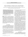
After a low brainstem stroke, a 73-year-old man experienced episodes of 90 degrees tilting of the visual environment in the sagittal plane evoked or terminated by voluntarily changing his head position. The episodes ceased 10 days after the stroke. This provocation by head position supports the idea that pathologic visual-vestibular interaction is at the basis of the room tilt illusion. |
| Subject |
Older people; Brain Stem, physiopathology; Cerebral Angiography; Head-Down Tilt; Humans; Illusions, physiology; Magnetic Resonance Imaging; Male; Perceptual Disorders, etiology; Stroke, complications; Stroke, pathology |
| Format |
application/pdf |
| Publication Type |
Journal Article |
| Collection |
Neuro-Ophthalmology Virtual Education Library: Journal of Neuro-Ophthalmology Archives: https://novel.utah.edu/jno/ |
| Publisher |
Lippincott, Williams & Wilkins |
| Holding Institution |
Spencer S. Eccles Health Sciences Library, University of Utah |
| Rights Management |
© North American Neuro-Ophthalmology Society |
| Setname |
ehsl_novel_jno |
| ID |
225591 |
| Reference URL |
https://collections.lib.utah.edu/ark:/87278/s6cr90fr/225591 |